 "
"
Team:Calgary/Project/BsDetector/SamplePreparation
From 2014.igem.org
| Line 3: | Line 3: | ||
<section class="Content Text Color-Normal"> | <section class="Content Text Color-Normal"> | ||
| - | + | <h2>Target Sequence Amplification</h2> | |
| - | <h2> | + | |
| - | + | ||
| - | + | ||
| - | + | ||
| - | + | ||
| - | + | ||
| - | + | ||
| - | + | ||
| - | + | ||
| - | + | ||
| - | + | ||
| - | + | ||
| - | + | ||
| - | + | ||
| - | + | ||
| - | + | ||
| - | + | ||
| - | + | ||
| - | + | ||
| - | + | ||
| - | + | ||
| - | + | ||
| - | + | ||
| - | + | ||
| - | + | ||
| - | + | ||
| - | + | ||
| - | + | ||
| - | + | ||
| - | + | ||
| - | <img src= | + | <p>A major advantage of our diagnostic system is an internal sample preparation stage, all that is required by the end-user is inputting the whole blood sample into the device. </p> |
| - | <p> | + | <p>Two technical challenges were identified regarding sample preparation, and amplification of our target DNA sequence. First, we must be capable of amplifying a target DNA sequence in a resource poor environment in addition to a potentially hostile climate. Conventional PCR methods are not feasible due to the inhibitory cost of thermocyclers, the necessary refrigeration of PCR components such as enzymes and buffers, and the training required to carry out and analyze PCR experiments. Secondly, we must be capable of amplifying the target DNA sequence directly from whole blood, a complex environment containing inhibitors of Taq polymerase.</p> |
| + | <p>Isothermal PCR (isoPCR) serves as an alternative to conventional PCR. isoPCR refers to the many molecular strategies that allow for amplification of a DNA sequence at a constant, relatively low temperature. Although there are several approaches to isoPCR, all strategies employ a molecular mechanism to circumvent the need for thermal denaturation of double-stranded DNA. In our system we utilized Recombinase Polymerase Amplification (RPA). RPA utilizes a strand-displacing polymerase to synthesize a complimentary strand of DNA using a double-stranded substrate; the recombinase and a single-stranded DNA-binding protein allow for oligonucleotide primers to stably bind to the template DNA. Because RPA is not dependent on a thermocylcer, it is capable of cycling through the amplification process continuously until the exhaustion of resources such as ATP and primers. Continuous amplification in RPA results in an amplified signal that is comparative to conventional PCR in a fraction of the time. For our purposes, in developing the sample preparation stage of our system, we used the commercially-available RPA kit purchased from TwistDx.</p> | ||
| + | <p>To eliminate the need for refrigeration, and further improve the shelf-life of our system, the components of isoPCR are freeze-dried, allowing activation by rehydration immediately prior to use. This was demonstrated to be effective as the TwistDx isoPCR kit provided us with freeze-dried pellets and rehydration buffer. </p> | ||
| + | <img src="https://static.igem.org/mediawiki/2014/7/7b/2014UCalgary_Freeze-dried_pellets.png" width="500" class="Center"> | ||
| + | <p class="Center"><b>Figure 1:</b> Freeze-dried pellets provided by the RPA Basic Kit from TwistDx. Pellets contain all components necessary for isoPCR, reaction is activated upon addition of hydration buffer and magnesium-acetate also provided in the kit.</p> | ||
| + | <p>Whole-blood compatibility in our amplification protocol was achieved using a Taq polymerase tolerant of inhibitors typically found in whole blood. Our sponsor KAPA provided us with KAPA Blood PCR Kits containing an engineered, second-generation Taq polymerase. By supplementing the RPA mixture with the blood-compatible Taq we achieved a greater signal output of isoPCR in the presence of blood. This demonstrated the efficacy of our system to amplify a target DNA sequence in the presence of blood, without the use of a thermocycler. </p> | ||
| + | <img src="https://static.igem.org/mediawiki/2014/9/96/2014UCalgary_Sept12_isoBloodHybrid_positiveCntrl_EDIT.png" width="500" class="Center"> | ||
| + | |||
| + | <p class="Center"><b>Figure 2:</b> Results of isoPCR using a 132bp positive control sequence provided with the RPA Basic Kit from TwistDx. Amplification was performed with blood concentrations of 0%, 1%, 5% or 10% v/v sheeps blood treated with the anti-coagulant citrate. Amplification was performed with either RPA exclusively, or with a 25% v/v substitution using the KAPA Blood PCR Kit mix containing the blood- compatible Taq polymerase. At 10% v/v blood, RPA failed to produce the expected amplicon; the addition of blood-compatible Taq produced the expected 132bp amplicon in the presence of 10% v/v blood.</p> | ||
| + | <p>Isolation of genomic DNA was not necessary for successful amplification of the target DNA sequence. Typically, conventional PCR requires a prolonged initial denaturation step (e.g. 10 minutes at 95C) to lyse the bacterial cells and release the DNA. However, our experiments demonstrate that isoPCR is effective on whole E. coli cells without prior lysis or preparation of template. </p> | ||
| + | |||
| + | |||
| + | <h2>Target Sequence</h2> | ||
| + | <p>To selectively detect the presence of N. meningititis we identified a unique yet conserved sequence present in all known N. meningititis genomes. The selected sequence was approximately 400bp, the maximum length of DNA our isoPCR was capable of amplifying. The specificity of our chosen sequence was confirmed by conventional PCR; a ~2.8kb amplicon was produced from the genomic DNA of six different N. meningititis strains, no amplicons were produced from the genomic DNA of five other pathogens including a commensal strain of Neisseria (Fig. 3). These results were replicated using isoPCR, amplifying a shorter target sequence of ~450bp. Although other smaller amplicons are produced by isoPCR these are the result of ‘primer noise’ inherent to the isoPCR method of RPA and may be reduced in the future by optimizing conditions such as primer concentration and screening primer variants for reduced primer noise (Fig. 4).</p> | ||
| + | <img src="https://static.igem.org/mediawiki/2014/1/1e/2014UCalgary_Sept10_MeningPrimer_SpecificityPCR_EDIT.png" width="500" class="Center"> | ||
| + | <p class="Center"><b>Figure 3:</b> Results of PCR on genomic DNA samples from eleven pathogenic parasites. All six N. meningititis strains produced the expected amplicon at ~2.8kb as indicated by white asterisks. The five other pathogens did not produce any products of the appropriate size.</p> | ||
| + | <img src="https://static.igem.org/mediawiki/2014/c/c0/2014UCalgary_Oct1_isoPCR_targetSeq400bp%28pure%29_EDIT.png" width="500" class="Center"> | ||
| + | <p class="Center"><b>Figure 4:</b> Results of isoPCR using RPA on the eleven genomic DNA samples as described in Fig. 3. Bands below 500bp are indicative of primer noise inherent to the RPA system. </p> | ||
| + | Alternative to PCR product DNA detection? | ||
| - | |||
| - | |||
| - | |||
| - | |||
Revision as of 17:42, 17 October 2014

Target Sequence Amplification
A major advantage of our diagnostic system is an internal sample preparation stage, all that is required by the end-user is inputting the whole blood sample into the device.
Two technical challenges were identified regarding sample preparation, and amplification of our target DNA sequence. First, we must be capable of amplifying a target DNA sequence in a resource poor environment in addition to a potentially hostile climate. Conventional PCR methods are not feasible due to the inhibitory cost of thermocyclers, the necessary refrigeration of PCR components such as enzymes and buffers, and the training required to carry out and analyze PCR experiments. Secondly, we must be capable of amplifying the target DNA sequence directly from whole blood, a complex environment containing inhibitors of Taq polymerase.
Isothermal PCR (isoPCR) serves as an alternative to conventional PCR. isoPCR refers to the many molecular strategies that allow for amplification of a DNA sequence at a constant, relatively low temperature. Although there are several approaches to isoPCR, all strategies employ a molecular mechanism to circumvent the need for thermal denaturation of double-stranded DNA. In our system we utilized Recombinase Polymerase Amplification (RPA). RPA utilizes a strand-displacing polymerase to synthesize a complimentary strand of DNA using a double-stranded substrate; the recombinase and a single-stranded DNA-binding protein allow for oligonucleotide primers to stably bind to the template DNA. Because RPA is not dependent on a thermocylcer, it is capable of cycling through the amplification process continuously until the exhaustion of resources such as ATP and primers. Continuous amplification in RPA results in an amplified signal that is comparative to conventional PCR in a fraction of the time. For our purposes, in developing the sample preparation stage of our system, we used the commercially-available RPA kit purchased from TwistDx.
To eliminate the need for refrigeration, and further improve the shelf-life of our system, the components of isoPCR are freeze-dried, allowing activation by rehydration immediately prior to use. This was demonstrated to be effective as the TwistDx isoPCR kit provided us with freeze-dried pellets and rehydration buffer.
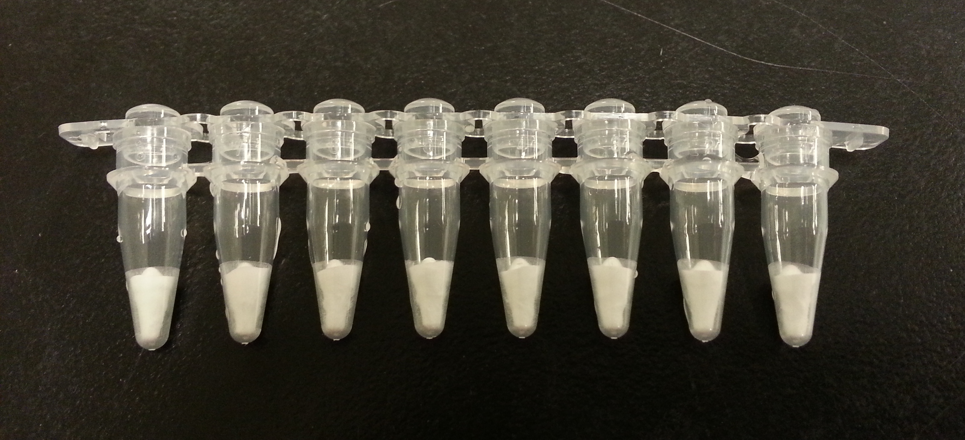
Figure 1: Freeze-dried pellets provided by the RPA Basic Kit from TwistDx. Pellets contain all components necessary for isoPCR, reaction is activated upon addition of hydration buffer and magnesium-acetate also provided in the kit.
Whole-blood compatibility in our amplification protocol was achieved using a Taq polymerase tolerant of inhibitors typically found in whole blood. Our sponsor KAPA provided us with KAPA Blood PCR Kits containing an engineered, second-generation Taq polymerase. By supplementing the RPA mixture with the blood-compatible Taq we achieved a greater signal output of isoPCR in the presence of blood. This demonstrated the efficacy of our system to amplify a target DNA sequence in the presence of blood, without the use of a thermocycler.

Figure 2: Results of isoPCR using a 132bp positive control sequence provided with the RPA Basic Kit from TwistDx. Amplification was performed with blood concentrations of 0%, 1%, 5% or 10% v/v sheeps blood treated with the anti-coagulant citrate. Amplification was performed with either RPA exclusively, or with a 25% v/v substitution using the KAPA Blood PCR Kit mix containing the blood- compatible Taq polymerase. At 10% v/v blood, RPA failed to produce the expected amplicon; the addition of blood-compatible Taq produced the expected 132bp amplicon in the presence of 10% v/v blood.
Isolation of genomic DNA was not necessary for successful amplification of the target DNA sequence. Typically, conventional PCR requires a prolonged initial denaturation step (e.g. 10 minutes at 95C) to lyse the bacterial cells and release the DNA. However, our experiments demonstrate that isoPCR is effective on whole E. coli cells without prior lysis or preparation of template.
Target Sequence
To selectively detect the presence of N. meningititis we identified a unique yet conserved sequence present in all known N. meningititis genomes. The selected sequence was approximately 400bp, the maximum length of DNA our isoPCR was capable of amplifying. The specificity of our chosen sequence was confirmed by conventional PCR; a ~2.8kb amplicon was produced from the genomic DNA of six different N. meningititis strains, no amplicons were produced from the genomic DNA of five other pathogens including a commensal strain of Neisseria (Fig. 3). These results were replicated using isoPCR, amplifying a shorter target sequence of ~450bp. Although other smaller amplicons are produced by isoPCR these are the result of ‘primer noise’ inherent to the isoPCR method of RPA and may be reduced in the future by optimizing conditions such as primer concentration and screening primer variants for reduced primer noise (Fig. 4).

Figure 3: Results of PCR on genomic DNA samples from eleven pathogenic parasites. All six N. meningititis strains produced the expected amplicon at ~2.8kb as indicated by white asterisks. The five other pathogens did not produce any products of the appropriate size.

Figure 4: Results of isoPCR using RPA on the eleven genomic DNA samples as described in Fig. 3. Bands below 500bp are indicative of primer noise inherent to the RPA system.
Alternative to PCR product DNA detection?