 "
"
Team:Freiburg/Content/Results/The combination
From 2014.igem.org
Mirja Harms (Talk | contribs) |
Mirja Harms (Talk | contribs) |
||
| Line 27: | Line 27: | ||
</figcaption> | </figcaption> | ||
</figure> | </figure> | ||
| - | + | ||
<div class="row category-row"> | <div class="row category-row"> | ||
| Line 40: | Line 40: | ||
</figcaption> | </figcaption> | ||
</figure> | </figure> | ||
| + | |||
| + | </div> | ||
<div class="col-sm-6"> | <div class="col-sm-6"> | ||
Revision as of 19:00, 17 October 2014
The combination
We, the iGEM Team Freiburg 2014, combined the spatial resolution of the light system with the specificity of our viral vector generating patterns in a homogenous cell culture.
Light induced receptor
Since our viral vector is specific for the mCAT-1 receptor that is under normal conditions only expressed in murine cells, we linked this specific receptor to the blue light system or the red light system and transfected non-murine cells with this receptor combinations. Mammalian cells which contain the inducible receptor expressed mCAT-1 after illumination. Cells expressing the receptor are visible by co-expression of mCherry. They were infected with our viral vector leading to EGFP expression. Cells of the same cell culture that were not illuminated were not able to express the receptor leading to no infection with our viral vector.
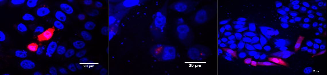
Red light induced receptor.
CHO cells were transfected with the red light system (PKM022) and the light induced receptor (p14rz_002). The receptor was labeled with mCherry; (left) after illumination with red light, (middle) incubation in the dark, (right) positive control. Cells were stained with DAPI. Labjournal

Blue light induced receptor.
HEK cells were transfected with the blue light system (PKM292 and PKM297) and the light induced receptor (p14ls_003). The receptor was labeled with mCherry. Cells were infected with MuLV EGFP afterwards; (left) incubation in the dark, (middle) after illumination with blue light, (right) cells expressing the light induced receptor were infected with MuLV. Labjournal

Blue light induced receptor in HEK293T
HEK cells were transfected with the blue light system (PKM292 and PKM297) and the light induced receptor (p14ls_003). The receptor was labeled with mCherry that was cleaved after expression, thus remaining in the cytoplasm. Cells were infected with MuLV EGFP afterwards; Overlay of all three channels (A); DAPI stained nuclei (B); EGFP expression in infected cells (C); mCherry expression in HEK293T cells since it was cleaved of the mCAT-1; (D) Objective plan apo 60x, 1.4 NA.
As we know that the transient mCAT-1 receptor that was not linked to a light system needs 24 hours for expression in HEK293 cells, we determined the expression time of the receptor that was induced by blue light after illumination for five hours. We used a receptor that was labeled with both, mCherry and an HA-tag (p14ls_003), for analysis with Western blot and fluorescence microscopy. The results show that the receptor had an expression peak at 24 hours after beginning of illumination.

Kinetics of the blue light induced receptor.
HEK cells were transfected with the blue light system (PKM292 and PKM297) and the light induced receptor (p14ls_003, mCherry labeled receptor). Pictures were taken after 12h, 15h, 18h and 24h. Labjournal
Pattern Generation
For generating pattern in homogenous cell layers we transfected HEK293T cells with the blue light system and the blue light induced receptor (p14ls_003). Dishes were covered with a photo mask preventing areas in the cell culture from light exposure. However, the blue light system is very sensitive to even low intensities of blue light. Due to scatterd light the receptor was activated in a huge area of the cell culture. Thus patterns were not visible to this time point due to by light scatter activated receptors (Fig.).

The light induced receptor was induced by light scatter.
HEK293T cells were transfected with the light system and the light induced receptor (p14ls_003) labeled with mCherry. Cells were illuminated using a photo mask that coverd parts of the cell culture and prevented them from light exposure. However, the receptor expression was activated.Labjournal