Team:Aachen/Project/2D Biosensor

From 2014.igem.org

(Difference between revisions)
m (Principle of Operation)
(Principle of Operation)
Line 57: Line 57:
<span class="anchor" id="biosensorpoo"></span>
-
''Cellock Holmes'' is devised based upon a SynBio approach comprised of a '''two-dimensional biosensor and a measurement unit'''. The two-dimensional biosensor is designed to recognize quorum sensing molecules secreted by the pathogen cells and to generate a distinct fluorescence signal, while the measurement device recognizes and analyses the produced signal. To overcome the limitation of our [https://2014.igem.org/Team:Aachen/Project/FRET_Reporter REACh construct] to bacteria that secrete quorum sensing molecules, we developed an alternative detection method ([https://2014.igem.org/Team:Aachen/Project/Gal3 Galectin-3 construct]), which is based on tagging cells with a fluorescent reporter.
+
''Cellock Holmes'' is devised based upon a SynBio approach comprised of a '''two-dimensional biosensor and a measurement unit'''. The two-dimensional biosensor is designed to recognize quorum sensing molecules secreted by the pathogen cells and to generate a distinct fluorescence signal, while the measurement device recognizes and analyzes the produced signal. On the molecular side, we use the '''[https://2014.igem.org/Team:Aachen/Project/FRET_Reporter REACh construct]''' to turn regular ''E. coli'' cells into ''Cellocks''. We also developed another molecular approach as an alternative to our REACh approach: The '''[https://2014.igem.org/Team:Aachen/Project/Gal3 Galectin-3 construct]''' overcomes the limitation of the REACh method to bacteria that secrete quorum sensing molecules because it is based on tagging cells with a fluorescent reporter.
[graph quorum senising]
[graph quorum senising]

Revision as of 07:46, 16 October 2014

2D Biosensor

With our 2D biosensor technology we are able to detect the pathogen Pseudomonas aeruginosa on solid surfaces. The sensor system is comprised of two distinct but inseparable modules, a biological part and a technical part:

  • Sensing chips containing Cellocks, our engineered detective cells that fluoresce in the presence of the pathogen, make up the biological part of Cellock Holmes.
  • Our measurement device WatsOn and the complementary softare Measurarty complete our sensing technology on the technical side.


Aachen 15-10-14 Principle of operation 2D sensor ipo.png

Principle of Operation

Cellock Holmes is devised based upon a SynBio approach comprised of a two-dimensional biosensor and a measurement unit. The two-dimensional biosensor is designed to recognize quorum sensing molecules secreted by the pathogen cells and to generate a distinct fluorescence signal, while the measurement device recognizes and analyzes the produced signal. On the molecular side, we use the REACh construct to turn regular E. coli cells into Cellocks. We also developed another molecular approach as an alternative to our REACh approach: The Galectin-3 construct overcomes the limitation of the REACh method to bacteria that secrete quorum sensing molecules because it is based on tagging cells with a fluorescent reporter.

[graph quorum senising]

Our sensor cells are immobilized in agar chips. To make the chips, we mix the sensing cells, also known as Cellocks, with liquid LB agar. In the course of our project, we designed a casting mold specifically for the production of our agar chips. When the agar has cooled down, the chips are cut out of the mold and are ready to use. Storage of the readily usable sensor chips is possible for 2 days at 4 °C when using LB medium or for 5 days if TB-medium is used. A detailed description of the sensor chip manufacturing can be found in our Protocols section.

Aachen 14-10-14 Flowsheet OD-device part1 ipo.png
Application of WatsOn for investigation of solid surfaces.


The application of Cellock Holmes for detection of P. aeruginosa cells is simple: fist, a sampling chip is placed on a hard surface that is potentially contaminated with the pathogen. Second, the sampling chip is removed from the surface and put onto one of our sensor chips. Theorectically the sensor chips could be directly used for sampling, but this was avoided in our project to match biosafety regulations. Third, the two layered chip-stack is put into a petri dish, which is inserted into our measurement device WatsOn for evalutation.


Aachen 14-10-14 Flowsheet OD-device part2 ipo.png
Mode of action inside WatsOn.

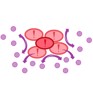
Inside WatsOn the chips are incubated at 37 °C and populations of microorganisms stuck on the sampling chip start to multiply. P. aeruginosa secrets an increasing number of quorum sensing molecules in growing cell colonies, which are taken up by our sensor cells and induce a fluorescence signal. In case of P. aeruginosa, we focused on quorum sensing molecules called N-3-oxo-dodecanoyl-L-homoserine lactones, or 3-oxo-C-12-HSLs for short, while our sensor cells contain of genetically modified E. coli cells which are able to elecit a fluorescent response to autoinducers. The 3-oxo-C-12-HSL detection system incorporated into the sensor cells is explained in more detail in the REACh Construct section.


Inside WatsOn' the chips can be illuminated with blue light at any time, whereby a picture of the chip is taken. The software Measurarty then analyzes any fluorescent signal.


Aachen 14-10-15 Medal Cellocks iNB.png

Achievements

Medium

Prior to using our own device for detection of fluorescence emitted by the sensor chips we used equipment readily available in the lab. A Molecular Imager® Gel DocTM XR+ from BIO-RAD was available which used UV and white light illuminators. Only two different filters were availble for the excitation ligth wavelength, which resulted in very limitted possibilities for excitation of fluorescent molecules. For example, it was possible to detect the expression of iLOV in our sensor chips, but in contrast detection of GFP was not possible. Thus the Gel DocTM XR+ was not ideal for our project. (iLOV_GFP_HM_1,5h.png)

Concerning the medium used for our sensor chips, LB medium showed a high background fluorescence when exposed to UV light. Surprinsingly the background fluorescence resulting from the LB medium was to high to detect a signal emitted by our sensor cells, thus we used minimal media (NA, M9, Hartman) in order to minimize background fluorescence. The appliction of minimal media was sufficient to minimize the background fluorescence, but this approach came with the drawback of minimal to zero growth of our sensor cells. (Chip_medium_geldoc.png)

Further experiments were conducted concerning long-time storage of the sensor chips. Storage at -20 °C resulted in the loss of our sensor cells. Adding 5-10% (v/v) glycerol ensured survival of the sensor cells, but resulted in an expression stop of fluorescence proteins. Thus the idea of long time storage of the sensor chips hab to be rejected. However, it was possible to store readily usable sensor chips for 2 days at 4 °C when using LB medium and storage for 5 days was possible, if TB medium was used. In our device, WatsOn, optimized wavelengths of 450 nm and 480 nm were used for excitation of iLOV and GFP, respectively. When exposed to both excitation wavelengths the LB medium showed minimal background fluorescence and no difficulties were observed in cultivation of our sensor cells. Further reduction of background fluorescence compared to LB medium was observed when using TB medium for sensor chip manufacturing in combination with fluorescence evaluation using the WatsOn device. (5Tage_K131026_neb_tb_1,5h)

Agar Concentration

For sensor chip manufacturing an optimal agarose concentration of 1.5% was found. When agarose concentrations below 1.5% (w/v) were used the sensor chips were easily damaged and were not transportable. Agar concentrations above 1.5% (w/v) had to be avoided, because the agarose started to solidify before it could be poured into the chip mold. Agarose was chosen above agar, because of more evely linkage and thus more homogenous chips. In addition agarose reduced diffusion of inducer molecules through the chip. Reduced diffusion was desired in order to achieve distinct fluorescent spots on the sensor chips.

Chip Form

Various approaches were tried for production of sensor chips with reproducable quality. The first approach was to cast every sensor chip indicidually. In order to archive a plain chip surface, which was required for high quality images, we tried to cast the sensor chips between two microscope slides. This approach had to be rejected, because the agar was too liquid. Second, we produced a closed mold in which the liquid agar was injected using a pipette, but we encountered a high frequency of bubbles in the chips when using this approach. Bubbles in the sensor chips resulted in problems during fluorescence evalutaion. (2_chipform.jpg)

Finally we used an open mold in which the agar was poured right after mixing with the sensor cells. When the agar was solidified the chips were cut out along precast immersions in the chip mold. An advantage of the open mold was the ability to simultaneously produce nine sensor chips whereby the surface tension of the liquid agar ensured a plane chip surface. (final_chipform.jpg)

Induction

For the induction of the used constructs we use IPTG or 3-oxo-C12 HSL. The sensor cells with K1319042 in BL21 can detect a IPTG concentration of 1 mM (0,2 µl). (2µl_IPTG_1mM_K1319042_1h.png) The sensor with the REACh constructs in BL21 can detect a IPTG concentration of ??? (Plate reader???) The sensor cells with K131026 in BL21 can detect an HSL concentration of 500 µg/ml (0,2 µl). It also can detect Pseudomonas aeruginosa. (Zeitaufnahmen bearbeitet von Arne)

Testing our Sensor Chips with a Platereader

300px
Testing K1319042 in our sensor chips
K1319042 in our sensorchip induced with 2 µl iPTG and measured with a Platereader. Blue color indicates no fluorescence, Red color indicates fluorescence.
300px
Testing K131026 in our sensor chips
K131026 in our sensorchip induced with 0.2 µl 3-oxo-C12 HSL and measured with a Platereader. Blue color indicates no fluorescence, Red color indicates fluorescence.

Detecting the 3-oxo-C12 HSL with K131026 in our sensor chip with WatsOn.

480px
Detection of 3-oxo-CC12 HSL with K131026
0,2 µl of 3-oxo-C12 HSL were put in the middle of the chip and then incubated at 37 °C in our WatsOn device.

Detecting Pseudomonas aeruginosa with K131026 in our sensor chip with WatsOn.

480px
Detection of Pseudomonas aeruginosa with K131026
Direct detection of Pseudomonas aeruginosa on our sensor chips. Sensor cell used were K131026.