Team:Aachen/Project/FRET Reporter

From 2014.igem.org

(Difference between revisions)
(References)
Line 156: Line 156:
== References ==
== References ==
 +
Broussard JA, Rappaz B, Webb DJ, Brown CM. Fluorescence resonance energy transfer microscopy as demonstrated by measuring the activation of the serine/threonine kinase Akt. Nat Protoc. 2013;8(2):265-81. doi:10.1038/nprot.2012.147.
 +
 +
{{Team:Aachen/Footer}}

Revision as of 14:42, 15 October 2014

The REACh Construct

On this page, we present our biosensor on the molecular level. Explore the different parts of our genetic device:


Aachen 14-10-13 GFP iNB.png

A Fluorescence Answer Faster Than Expression

Biosensors often work with a system that is comprised of a reported gene under the control of a promoter that is induced directly by the chemical that the sensor is supposed to detect. In the case of our 2D biosensor for Pseudomonas aeruginosa, the expression of our reporter gene, GFP, would be directly induced by the activity of the bacterium's quorum sensing molecules. However, transcription, translation, folding and post-translational modifications take their time. Since our goal is to detect the pathogen as fast as possible, we wanted to use a system that gives a fluorescent answer fast than just expressing the fluorescent protein.

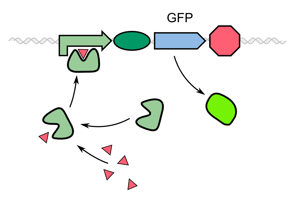
Aachen Traditional biosensor.png
Approach of a traditional biosensor
In this model, the expression of GFP is directly controlled by a promoter whose activator binds to a molecule secreted by the pathogen.

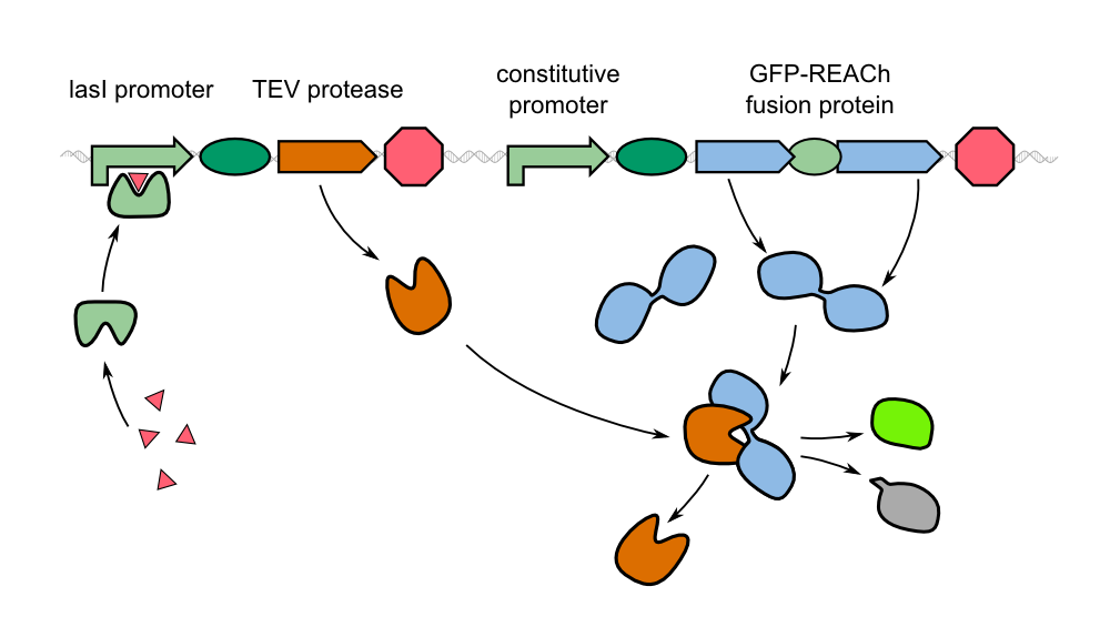
Instead of the traditional approach, we constitutively express our reporter gene in its quenched form. As GFP_REACh fusion protein, fluorescence is suppressed. Our biosensor gives a response when homoserine lactones of Pseudomonas aeruginosa are taken up by our sensor cells where the autoinducer activates the expression of the TEV protease by binding to the LasI promoter in front of the protease gene.

Aachen REACh approach.png
Our novel biosensor approach
Expression of the TEV protease is induced by HSL. The protease cleaves the GFP-REACh fusion protein to elicit a fluorescence response.

This approach has two advantages:

  • When Pseudomonas aeruginosa is detected by our cells, the reporter protein is already expressed and only waits to be cleaved off the REACh quencher. The cleavage reaction catalyzed by the TEV protease is a faster process than expression and correct folding of GFP. This way we hope for an earlier response by our sensor cells.
  • While a certain concentration of homoserine lactone will produce the same number of gene read-outs, one TEV protease can cleave many GFP_REACh constructs. Through the cleavage step we therefore introduce an amplification step into our system. With the TEV protease, we will be able to produce a much stronger signal in a short time interval.

Add: [Diagram of the expression levels/signal strength over time]

Aachen 14-10-13 FRET Arrows iNB.png

The FRET (Förster Resonance Energy Transfer) System

Förster resonance energy transfer (FRET), sometimes also called fluorescence resonance energy transfer, is a physical process of energy transfer. In FRET, the energy of a donor chromophore, whose electrons are in an excited state, is passed to a second chromophore, the acceptor. The energy is transferred without radiation and is therefore not exchanged via emission and absorption of photons. The acceptor then releases the energy received from the donor, for example, as light of a longer wavelength.

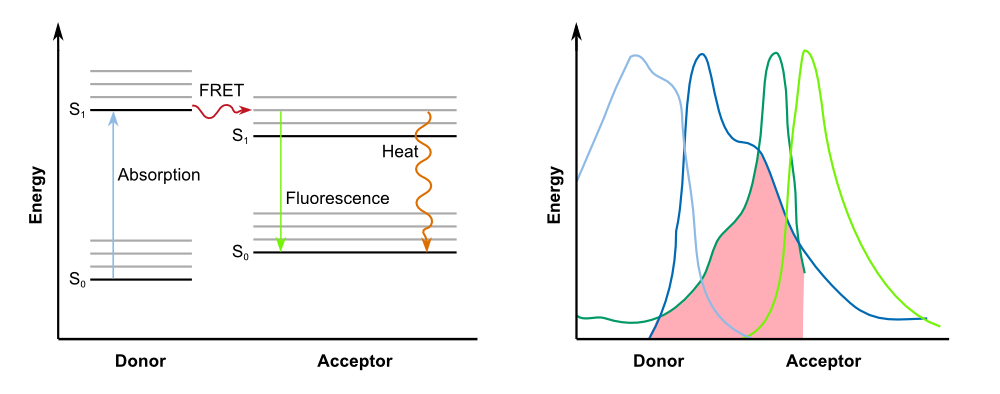
In biochemistry and cell biology, fluorescent dyes, which interact via FRET, are applied as "optical nano metering rules", because the intensity of the transfer is dependend on the spacing between donor and acceptor, and can be observed up to distances of 10 nm. This way, protein-protein interactions and conformational changes of a variety of tagged biomolecules can be observed. For efficient FRET to occur, there must be a substantial overlap between the donor fluorescence emission spectrum and the acceptor fluorescence excitation (or absorption) spectrum ([http://www.nature.com/nprot/journal/v8/n2/full/nprot.2012.147.html Broussard et al., 2013]), as described in Fig.2.

Aachen 14-10-07 Jablonski Diagram and Absorption Spectra iNB.png
FRET betweeen donor and acceptor
On the left: Jablonski diagram showing the transfer of energy between donor and acceptor; on the right: For successful FRET, the emission spectrum of of the donor has to overlap with the absorption spectrum of the acceptor.

However, fluorescence is not an essential requirement for FRET. This type of energy transfer can also be observed between donors that are capable of other forms of radiation, such as phosphorescence, bioluminescence or chemiluminescence, and fit acceptors. Acceptor chromophores do not necessarily emit the energy in form of light, and can lead to quenching instead. Thus, this kind of acceptors are also referred to as dark quenchers. In our project, we use a FRET system with a dark quencher, namely our REACh construct.

Aachen 14-10-13 REACh iNB.png

REACh Proteins - Dark Quenchers of GFP

In 2006, [http://www.pnas.org/content/103/11/4089.full Ganesan et al.] were the first to present a previously undescribed FRET acceptor, a non-fluorescent yellow fluorescent protein (YFP) mutant called REACh (for Resonance Energy-Accepting Chromoprotein). YFP can be used as a FRET acceptor in combination with GFP as the donor in FRET microscopy and miscellaneous assays in molecular biology. The ideal FRET couple should possess a large spectral overlap between donor emission and acceptor absorption - as illustrated in Fig. 2 - but have separated emission spectra to allow their selective imaging.

To optimize the spectral overlap of this FRET pair, the group obtained a genetically modified YFP acceptor. Mutations of amino acid residues that stabilize the excited state of the chromophore in enhanced YFP (EYFP) resulted in a non-fluorescent chromoprotein. Two mutations, H148V and Y145W, reduced the fluorescence emission by 82 and 98 %, respectively. Ganesan et al. chose the Y145W mutant and the Y145W/H148V double mutant as FRET acceptors, and named them REACh1 and REACh2, respectively. Both REACh1 and REACh2 act as dark quenchers of GFP.

256px
Homology model of REACh.

Aachen 14-10-13 Fusion Protein iNB.png

Producing a GFP-REACh Fusion Protein

In our project, we reproduced the REACh1 and REACh2 proteins by subjecting an RFC-25 compatible version of the BioBrick [http://parts.igem.org/Part:BBa_E0030 E0030] (EYFP) to a QuikChange mutation, creating the BioBricks K1319001 and K1319002, respectively. Subsequently, we fused each REACh protein with GFP (mut3b) which is available as BioBrick [http://parts.igem.org/Part:BBa_E0040 E0040]. The protein complex was linked via a protease cleavage site, K1319016. As constitutive promoter we use [http://parts.igem.org/Part:BBa_J23101 J232101]. When GFP is connected to either REACh quencher, GFP will absorb light but the energy will be transferred to REACh via FRET and then emitted in the form of heat; the fluorescence is quenched. Our cells also constitutively express the [http://parts.igem.org/Part:BBa_C0179 LasR] activator. Together with the HSL molecules from P. aeruginosa, LasR binds to the [http://parts.igem.org/Part:BBa_J64010 LasI] promoter that controls the expression of the TEV protease, which we make available as K1319004. When the fusion protein is cleaved by the TEV protease, REACh will be separated from GFP. The latter will then be able to absorb and emit light as usual.

Aachen 14-10-08 REACh approach with BioBricks iNB.png
Composition of our biosensor
For our biosensor, we use a mix of already available and self-constructed BioBricks.


Aachen 14-10-13 TEV Protease iNB.png

Cutting the Fusion Protein with the TEV Protease

To cut the GFP_REACh fusion protein off, we chose Tobacco Etch Virus (TEV) protease, a highly sequence-specific cysteine protease, that is frequently used for the controlled cleavage of fusion proteins in vitro and in vivo. The native protease also contains an internal self-cleavage site. This site is slowly cleaved to inactivate the enzyme. The physiological reason for the self-cleavage is unknown, however, undesired for our use. Therefore, our team uses a variant of the native TEV protease that contains the mutation S219V which results in an alteration of the cleavage site so that self-inactivation is diminished.

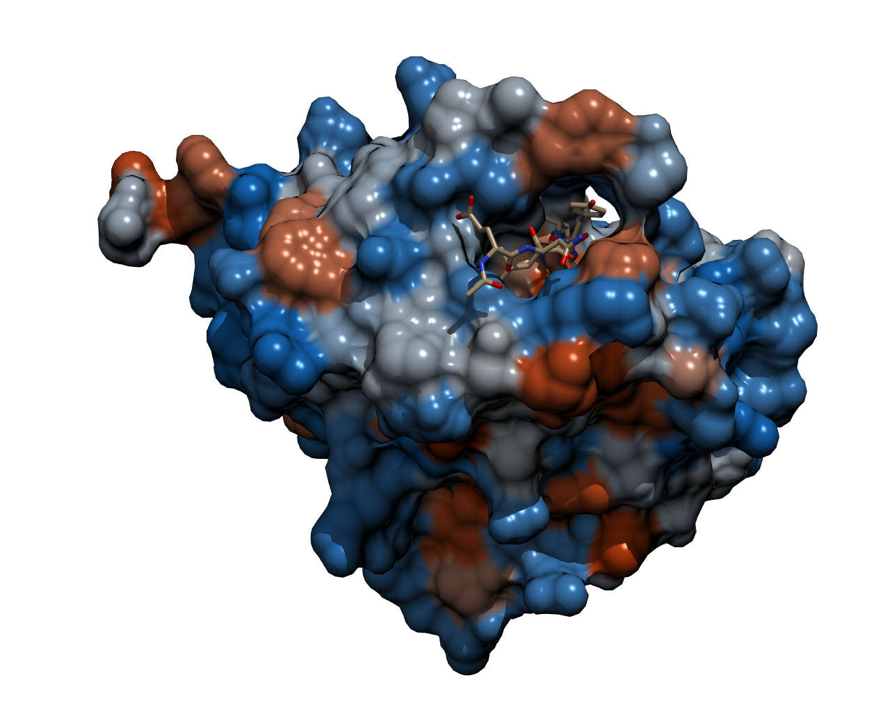
Aachen TEV Protease Model.png
TEV protease with a bound peptide
This picture shows the TEV protease with a peptide chain bound in the binding pocket ready to be cleaved. The bound peptide chain has the recognition sequence inside the binding pocket.

Though quite popular in molecular biology, the TEV protease is not avaiable as a BioBrick yet. Hence, the Aachen team introduces a protease with anti-self cleavage mutation S219V and codon optimized for E. coli [http://parts.igem.org/Part:BBa_K1319004 to the Parts Registry this year.]

Aachen 14-10-15 Medal Cellocks iNB.png

Achievements

Awaiting synthesis...

References

Broussard JA, Rappaz B, Webb DJ, Brown CM. Fluorescence resonance energy transfer microscopy as demonstrated by measuring the activation of the serine/threonine kinase Akt. Nat Protoc. 2013;8(2):265-81. doi:10.1038/nprot.2012.147.