Team:LMU-Munich/Project/BioBrickBox

From 2014.igem.org


Bacillus BioBrickBox

In 2012, the iGEM-team LMU-Munich began the task to develop essential BioBricks, like vectors, promoters, reporters and affinity tags, especially suited for the use in Bacillus subtilis in order to establish a new chassis in the Escherichia coli-dominated world of iGEM. Even after the finals of the iGEM competition in 2012, the project was pursued further since its importance for synthetic biology and resulted in a publication in the Journal of Biological Engineering about this so called Bacillus BioBrick Box.

This year, our team aims to enhance the Bacillus BioBrick Box by adding new parts, like different colored fluorescent proteins, a RBS-collection, evaluating protein linkers for B. subtilis and creating a redesigned backbone for resistance free strain generation.

Resistance free strain generation

With BaKillus as an medical application it is most likely that it will be released - though in small amounts - into the environment. Therefore we sought after a solution in order to keep the final BaKillus free of unnecessary antibiotic resistances. In order to do so we developed a resistance free gene insertion strategy for integrative Bacillus subtilis vectors.

The upp gene from Bacillus subtilis W168 encodes for a Uracilphosphoribosyl transferase (UPRTase). Its key reaction in uracil salvage is the reaction of a uracil molecule with a 5'-phosphoribosyl-a-1- pyrophosphate (PRPP) molecule, resulting in the formation of UMP. A second locus, the pyrR gene, encoding a second UPRTase has been identified. However it has been shown, that the UPRTase derived from pyrR locus has an influence on overall UPRTase activity of < 1 %. [1] This makes the upp-derived UPRTase the only physiologically relevant catalyst for UPRTase activity. Exposure to the pyrimidine analogue 5-Fluorouracil UPRTase results in production of 5-fluoro-dUMP, a very potent inhibitor of the thymidylate synthase (Neuhard, J. (1983) Utilization of preformed pyrimidine bases and nucleosides. In Metabolism of Nucleotides, Nucleosides and Nucleobases in Microorganisms. Munch- Petersen, A. (eds). New York: Academic Press, pp. 95– 148. ). As a result 5-FU is toxic to the Bacillus subtilis W168 strain. B. subtilis 5FU-resistant (5FUR) mutants selected on low drug concentration (10 mM 5FU) are UPRTase-defective. [2]. This has made upp a go to choice for negative selection in combination with an B. subtilis W168 Δupp strain. So far it has been used to make clean in-frame deletions and point mutations. [3]

However, to our knowledge, no application using upp for clean insertions has been established so far.

The I-SceI Restricition endonuclease has a highly specific recognition sequence of 18 nucleotides. No such sequence is present in the B.subtilis W168 strain. It creates a double strand break at targeted location, which leads to an increased rate of repair at the specific site. By this, the rate of homologous recombination is increased by a factor of 100. [4]

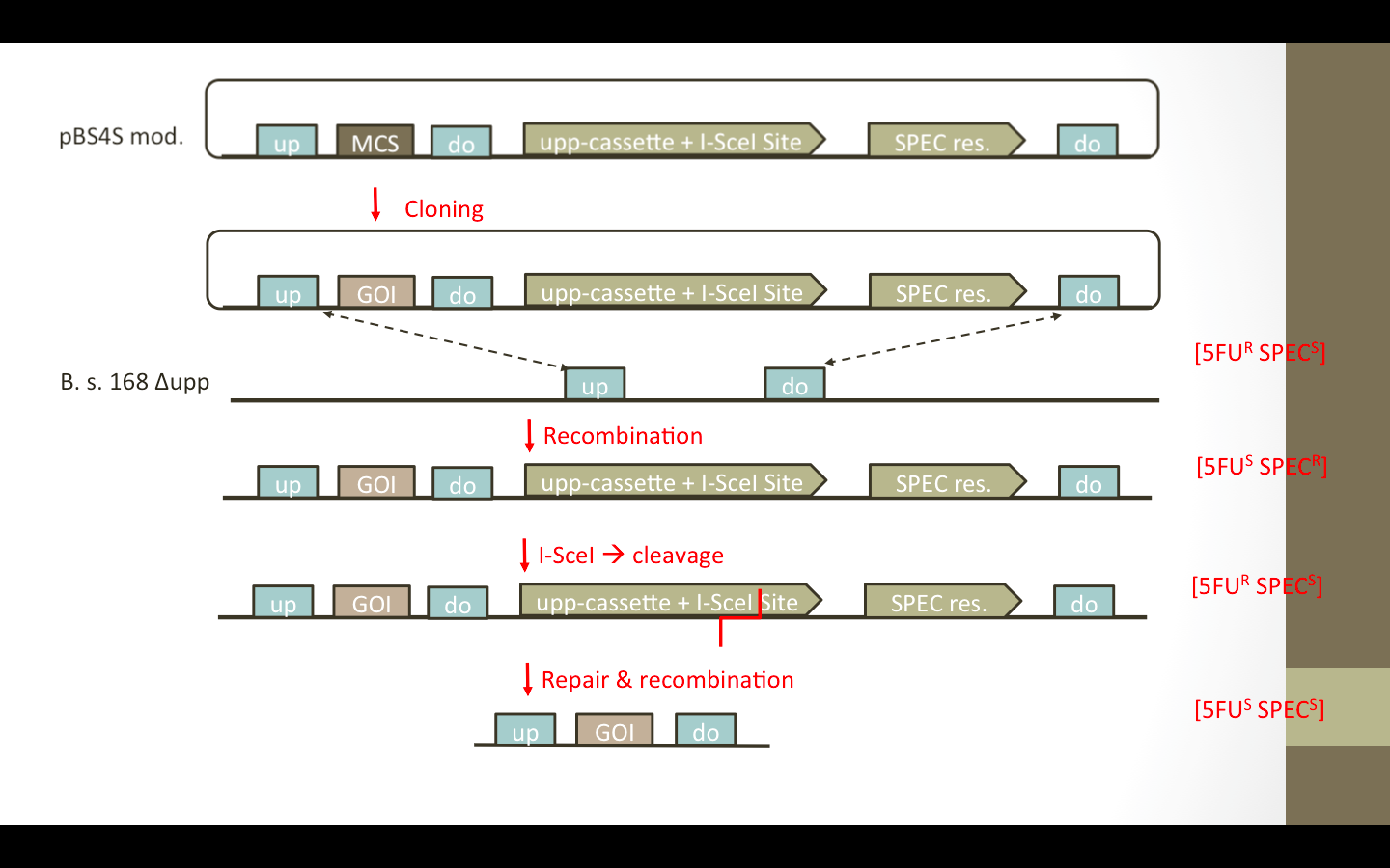
The Plan was to restructure the BioBrick compatible, integrative Bacillus subtilis vectors pSB1C, pSB2E and pSB4S in a fashion that leads to deletion of the antibiotic resistance after an insertion of a gene of interest has been generated. Integration of those basic vectors is achieved via homologous recombination between the amyE/lacA/thrC locus, respectively, and the corresponding up and down fragments on the vector. This leads to an insertion of the gene of interest within the RCF10 compatible multiple cloning site and the resistance for positive selection (Cmr/MLSr/Specr).

We wanted to add the upp-cassette, containing an I-SceI site, as well as an additional up/down fragment to the vector. The desired vectors are presented in Fig. 1. The upp-cassette will allow negative selection in 5FU media. The contained I-SceI site will be cut by the I-SceI restriction endonuclease which is encoded on a helper plasmid pEBS-cop1. [5] (Figure)


pEBS-cop1. Plasmid containing the i-sceI gene

Cloning Strategy

LMU2014 BioBrickBox ResistanceFree Cloning Strategy.png

Lab work on this project only started at end of July So far the upp cassete has been modified to fullfill the Freiburg Standard (RCF25) criteria and was successfully sequencend and submitted to the registry as BioBrick part BBa_K1351023. Creation of the complete and functional backbones has not been successfull. We assume that due to the duplication of homologous sequences for the integration into Bacillus subtilis genomic loci a deletion has occured during phusion PCR. This is supported by the results of multiple digests of the resulting plasmid by different restriction enzymes.

We plan on continuing work on this project after the iGEM Competition

Fluorescent Proteins

Fluorescent proteins (FPs) are important tools in research on a cellular or molecular basis and in the past years, many different FPs with very diverse qualities related to color, brightness, duration, bleaching, maturation time and other parameters were developed. Unfortunately, most of these FPs are optimized for use in E. coli, yeast or mammalian cells, and we aim to evaluate the properties of different FPs for the use in B. subtilis to enhance our Bacillus BioBrick Box even further.

Fluorescent Proteins are often derivates of the first described FP, GFP, which was isolated from the jellyfish Aequorea victoria. This protein is of typical barrel-shape, built from 11 ß-sheets and a chromophore in the middle of the barrel. This barrel structure is typical for fluorescent proteins, whether they derive from GFP or not. Other colors than green were developed by changing single amino acids in the chromophore of GFP (e.g.: Yellow: YFP and Cyan: CFP) or by analyzing fluorescent proteins from other organisms, like Discosoma striata, a coral, from which some of our red FPs, like dsRed derive. FPs are an important tool in today’s research, for example in order to study gene expression or the location of specific proteins in a cell or a whole organism. This is the reason, why we tried to establish some different colors of FPs for the usilization in B. subtilis by evaluating them for the Bacillus BioBrick Box.
Seven different FPs (Table 1) where chosen and either obtained from the registry or from the lab group Mascher. The BioBrick <partinfo>BBa_E1010</partinfo>was mutated via site directed mutagenesis by overlap extension PCR in order to delete two AgeI-Restriction sites and make the BioBrick compatible for the Freiburg standard RFC25. This improved BioBrick is called BBa_K1351021 The BioBricks and BBa_K592100 (mTagBFP) where both provided with the necessary overhangs for the Freiburg standard, the other FPs where already provided with the proper restriction overhangs. These seven FPs where combined C- and N-terminally with a His-Tag (BBa_K823037) and cloned into the vector pBS0K-Pspac (BBa_K1351040) and transformed into B. subtilis.


Table 1: Used fluorescent proteins
BioBrick number Name Color Excitation Peak Emission Peak
BBa_K1351021 DsRed Red 584nm 607nm [1]
BBa_K592100 mTagBFP Blue 399nm 456nm
BBa_K1159302 eCFP Cyan 439nm 476nm
BBa_K1159301 sYFP2 Yellow 515nm 527nm
BBa_K823039 gfpmut1 Green 395nm 509nm
BBa_K1351042 mcherry Red 587nm 610nm
BBa_K823029 mKate 2 Red 588nm 633nm


LMU14 FP Gesamtkonstrukt.png

Fluorescence of GFP under expression of promoter Pspac

The mutagenesis of the Biobrick E1010 was successful and confirmed by sequencing, the thus created Biobrick is called BBa_K1351021. The fusion with the His tags were also successfully conducted and confirmed by sequencing. The ligation into the vector pBS0K-Pspac was confirmed via Colony PCR and the transformation into B. subtilis was conducted successfully.

The intensity and spectrum of the fluorescence was measured by a TECAN Plate Reader, kindly provided by the AG Leonhardt from the LMU.

The fluorescence of all of the proteins, however, proved to be not very good and often at the same level as the auto fluorescence of the B. subtilis wild type. The examination of the cells under a microscope revealed, that the promoter is not suited for this experimental set up, since the gene expression is very heterogeneous and rather low.


This problem will be solved by recloning the FPs into the vector pBS1C (BBa_K823023) together with the xylose-inducible promoter Pxyl(BBa_K1351039).

Linkers

Linkers are short peptides used to fuse two protein domains (or even whole proteins). They are designed not to interact with the fused parts. A linker of the wrong length can lead to sterically inactivated (if the linker is too short) or instable (if it is too long) constructs. Last but not least, linkers need to exhibit a certain flexibility. We evaluated a set of linkers partially already found in the registry using FRET.

Förster Resonance Energy Transfer (FRET) is a phenomenon that occurs when two fluorophores with overlapping spectra are not more than 10 nanometers apart. When the donor fluorophore gets excited, and FRET happens, it does not emit all the energy as light, it transferres a part of the energy non-radiatively to the acceptor fluorophore, which starts fluorescing. The efficiency of this energy transfer decays with the sixth power of the distance between donor and acceptor. This makes FRET an ideal tool to measure or verify very short distances, like the ones between interacting proteins in vivo. A common application would be tagging two supposedely interacting proteins with the FRET couple and measuring acceptor fluorescence. Obviously this technique lends itself well to the evaluation of linkers.

mTurquoise (donor) and mNeonGreen (acceptor) were used as FRET-couple for this study. Translational fusion constructs were created according to a modified RFC25 standard. From the registry we took the linkers BBa_K243004, BBa_K243005, BBa_K243006, BBa_K157009, BBa_K157013, BBa_K243029 and BBa_K243030. We also used this method to characterize our own linkers BBa_K1351009 and BBa_K1351035. For each linker a construct with mTurquise fused to it N-terminally and mNeonGreen C-terminally was created, as well as a construct where the FPs were oriented the other way around.

The constructs have been created, and fluorescence has been observed. Due to time concerns, the FRET-assay has not yet been performed.

Bacillus RBS collection

Ribosome Binding Sites (RBS) are essential for gene expression, since they are required for translation initiation. Much to our dismay, there used to be no Bacillus RBS in the registry. So we designed a broad-range RBS library for B. subtilis, aiming for maximum translation initiation rate space coverage.


RBS are short nucleotide sequences that are complemetary to the 3' end of 16S rRNA, and thus are bound by the ribosome during translation initiation. The RBS is usually found ~8 nucleotides upstream of the start codon in procaryotes. In B. subtilis the perfect consensus RBS is AAGGAGGGATA (Bba_K1351028).
Using the Salis lab's RBS calculator
we calculated our RBS library with the sequence ARRRRRRGATA. The 7 RBS from this collection that were submitted as BioBricks (BBa_K1351028-BBa_K1351034) are the ones that offer the best coverage of translation initiation rate space, number 1 being the optimal Bacillus RBS and number 7 being the weakest.
The constructs have been created, the evaluation however is not yet finished.


Hi there!

Welcome to our Wiki! I'm BaKillus, the pathogen-hunting microbe, and I'll guide you on this tour through our project. If you want to learn more about a specific step, you can simply close the tour and come back to it anytime you like. So let's start!

What's the problem?

First of all, what am I doing here? The problem is, pathogenic bacteria all around the world are becoming more and more resistant against antimicrobial drugs. One major reason for the trend is the inappropriate use of drugs. With my BaKillus super powers, I want to reduce this misuse and thus do my part to save global health.

Sensing of pathogens

To combat the pathogenic bacteria, I simply eavesdrop on their communication. Bacteria talk with each other via quorum sensing systems, which I use to detect them and trigger my responses.

Adhesion

The more specific and effective I can use my powers, the lower the danger is of provoking new resistance development. So I catch pathogens whenever I get hold of them and stick to them until my work is done.

Killing

Talking about my work - killing pathogens is finally what I am made for. In response to quorum sensing molecules of the pathogens, I export a range of antimicrobial substances leading to dissipation of biofilms and the killing of the targeted bacteria.

Suicide switch

When the job is done and all the bad guys are finished, you don't need a super hero anymore. So after fulfilling my work I say goodbye to the world by activating my suicide switch.

Application

Of course I'm not only a fictional hero, but a very real one. In two different prototypes, I could be used for diagnosis or treatment of pathogen-caused diseases. However, there is still a whole lot of regulational and economical questions that have to be answered before.

See you!

So now you know my short story - and it is time for me to return to my fight for a safer world. Feel free to take a closer look on my super powers, the process of my development or the plans for a medical application.