 "
"
Team:Freiburg/Content/Results/Vector
From 2014.igem.org
Mirja Harms (Talk | contribs) |
Mirja Harms (Talk | contribs) |
||
| Line 23: | Line 23: | ||
</div> | </div> | ||
| - | < | + | <center> |
<ul> | <ul> | ||
<li>specificity | <li>specificity | ||
| Line 29: | Line 29: | ||
<li>life time | <li>life time | ||
<li>capacity of stable integrate genes | <li>capacity of stable integrate genes | ||
| - | </ul></ | + | </ul> |
| + | </center> | ||
<h2>1.1 Specificity of MuLV</h2> | <h2>1.1 Specificity of MuLV</h2> | ||
Revision as of 10:36, 3 October 2014
1 The Vector
Our system provides a possibility to stable transfer genes into the genome of cells in spacial and temporal resolution. This transfer is carried out by a vector derived from a murine leukemia virus. Since the vector is one essential part of the system its characterisation is important. The virus is specific for a receptor that is only expressed in mouse cells. For the purpose of our system, this specificity offers the opportunity for targeting only these cells we want to be infected. Therefore, important aspects the vector must provide are:

- specificity
- high efficiency
- life time
- capacity of stable integrate genes
1.1 Specificity of MuLV
An important aspect for the function of our system as well as for its safety is the specificity of the vector regarding infection of different kind of cells. The vector deriving from the murine leukemia virus is specific for cells carrying the mouse specific CAT-1. Cells that do not have this specific receptor are not targeted by the vector. In order to test the specificity of the system, different kind of cells were incubated with the vector containing EGFP. Since EGFP is stable integrated by the system, infected cells are identified by a green fluorescence that was analysed via flow cytometry (figure 1).
We tested two human cell lines, human embryonic kidney cells as well as human lung epithel carcinoma cells, for their capability of being targeted by the vector. In addition, mouse fibroblasts that express the mouse specific CAT-1 receptor were tested for a positive control. As shown in figure 1 both human cell lines did not express EGFP after incubation with the vector indicating that they were not targeted. However, many cells of the mouse cell line were expressing EGFP after infection.

Fig.1: Description of the experiment (as picture)

Fig.X: FACS analysis of different cell lines incubated with the CAT-1 specific viral vector
Left: Different cell lines not incubated with the vector as negative control, Middle: different cell lines infected with MuLV IRES EGFP (50% in completed growth medium), Right: cells transfected with the mouse specific receptor CAT-1 (rz006) were infected with the viral vector. The infection efficiency is directly correlated with transfection efficiency of the receptor into the different kind of cells.
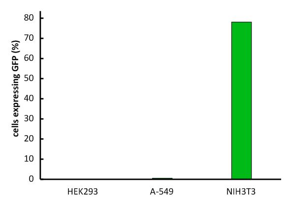
Fig.1: Infection of different cell lines with the viral vector derived from the murine leukemia virus.
In order to investigate the specificity of the viral vector, two human cell lines, human embryonic kidney cells as well as lung epithel carcinoma cells, were infected and the percentage of cells expressing EGFP was analyzed by flow cytometry.
The vector derived from the murine leukemia virus is specific for the murine CAT-1 receptor. Therefore it is not able to infect human cell lines!
1.2 Optimization of transduction
An importang aspect of our system is the infection rate of the viral vector. Therefore, we tried to optimize our transduction protocol reaching almost 100% efficiency in murine cell lines like NIH3T3 cells. capability from 16% at the beginning to almost 100%.
Fig.: nice pictures of nice cells.
Fig.: Transduction efficiency of the viral vector in mouse cells.
With the viral vector used in our system transduction efficiency in murine cells was optimized to almost 100%!
1.3 Expression of different viral constructs

Fig.: pMIG constructs
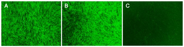
Fig.: Transduction efficiency of the viral vector in mouse cells.
bla bla
1.4 Virus dilution and concentration

Fig.: Concentrated virus
blubb!