 "
"
Team:Freiburg/Content/Results/Receptor
From 2014.igem.org
The Receptor
The specificity of our system is based on the murine CAT-1 receptor which serves as the viral entry site of our vector into target cells. We could show that our viral vector system can be used to stably integrate genes into the genome of murine cells. We could also target any other cell line by transferring the gene for the murine CAT-1 receptor in these cells before transduction. Since mCAT-1 is naturally only present in murine cell lines, we use human cell lines for transfection of the receptor gene. Expression of a reporter demonstrates that only the cells expressing the mCAT-1 receptor are infected by the viral particles, thus making particular gene transfer possible.
Localisation Receptor
Since, the mCAT-1 receptor serves as the entry site of our viral vector, it is essential that it is expressed on the surface of target cells. In order to determinate the localization of the mCAT-1 receptor, we labeled the C-terminus of the mCAT-1 with the fluorescence protein mCherry and transfected this construct in human embryonic kidney (HEK293) cells. After distinct time points cells were imaged with a confocal scanning laser microscope. The mCAT1-mCherry was found predominantly at the surface of the cells.
Receptor Functionality
Transduction of genes into murine cell lines which naturally express the mCAT-1 receptor occurred with an efficiency over 80%. For our system we need a functional expression of the receptor in non-murine cell lines, i.e. the receptor has to serve as an entry site for the virus, and the virus has to efficiently deliver its cargo into the target cell. We tested the functionality of the mCAT-1 receptor by infecting HEK293 cells expressing the receptor with the virus containing EGFP as a cargo. The presence of green fluorescent cells in the infected cultures of different non-murine cell lines indicates that the receptor is not only expressed but can be also used as an entry site by the virus.
During our projekt we generated different receptor constructs all of them labeled with differnt markers. We tested all of them for their functionality (Fig.). Analysis with flow cytometry show that all cells that were transduced by the viral particles transferring EGFP also express mCherry, i.e. only cells expressing the receptor were infected.

Figure : HEK293 cells transfected with different receptor constructs and infected with MuLV EGFP afterwards.
The cells were transfected with pQCXIN (original vector containing SLC7A1) (A), p14rz_004 (labeled with HA-tag)(B) and p14rz_006 (labeled with HA-tag and mCherry)(C). Cells were transduced with MuLV after 24 hours. Pictures were taken after 48 hours.
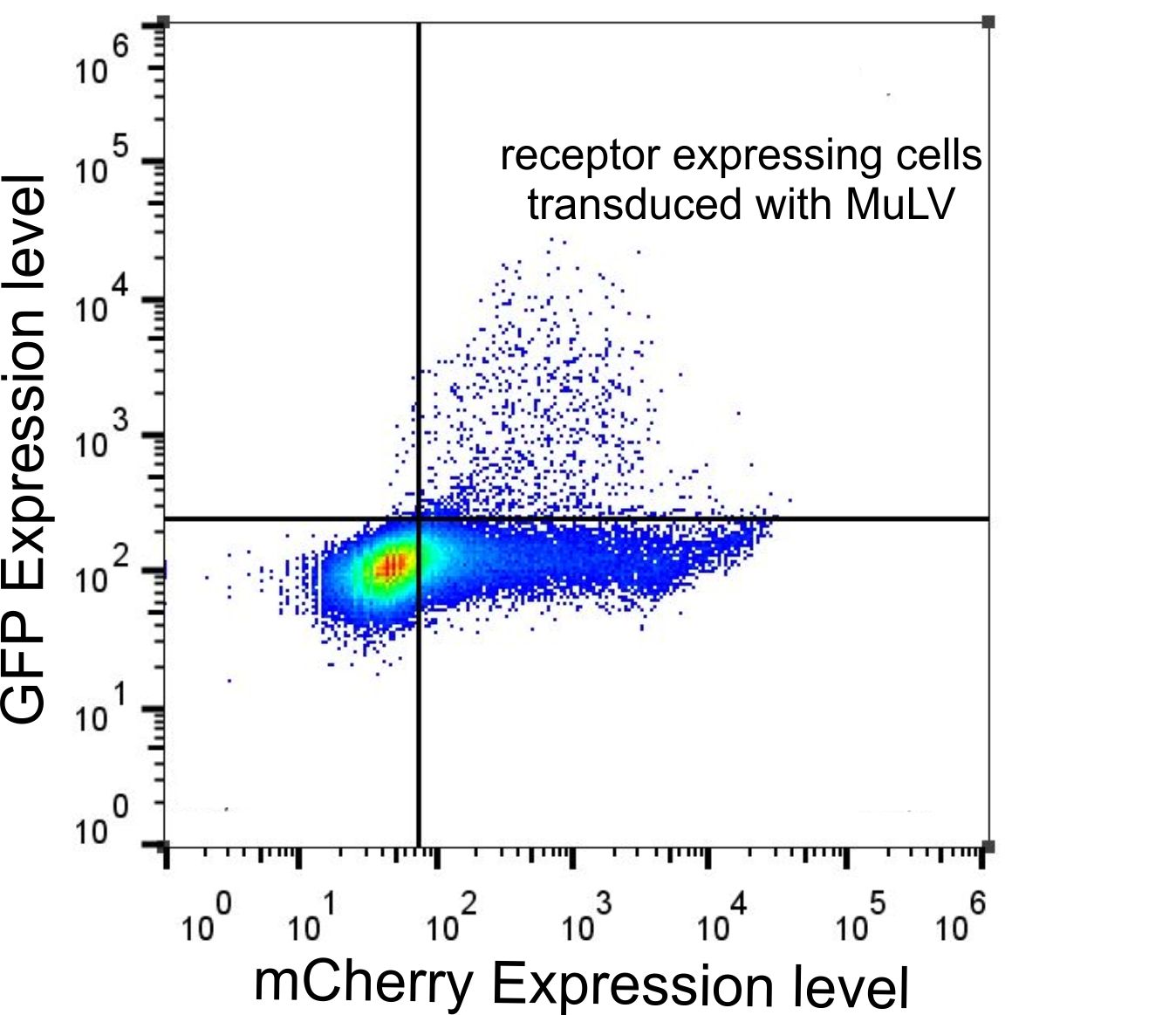
FACS data of HEK293 cells tranfected with the receptor and transduced with MuLV EGFP.
HEK cells were transfected with the p14rz_006 that was labeled with mCherry and infected with MuLV transferring EGFP. Cells were analyzed with flow cytometry 48 hours after transduction.
Receptor Expression
The time point for viral infection was adjusted to the time the receptor needs for expression in target cells. In order to determine the expression time of mCAT-1 in HEK293 cells, we transfected the cells with the HA-tagged mCAT-1 (p14rz_006). Cells transfected with receptor DNA were analyzed after different incubation times. As the expression of the receptor peaks at 24h after transfection, we used this time point for viral infections.

Figure 1: Comparing the blue light system in CHO cells and in HEK293 cells and comparison to the red light system in CHO cells.
To test the efficiency of the light system light induced SEAP was used as a marker. A SEAP assay was performed after 24 hours of incubation.

Efficiency of the blue light system using different durations of illumination.
To test the efficiency of the light system light induced SEAP was used as a marker. A SEAP assay was performed after 24 hours of incubation. Cells were incubated with blue light for 1 hour, 2.5 hours and 5 hours.