Project
・Back graund
・Link Light Assay
・HRV protease
・Methodology
Back graund
the secondary immune response
As a person's immune system encounters foreign substances (antigens), the components of acquired immunity learn the best way to attack each antigen and begin to develop a memory for that antigen.
As for the immune system, the antigen presenting cell (APC) such as the dendritic cell begins in the place where an antigen is taken in by an endocytosis.
The antigen taken in is broken down to peptide by lysosome.
The antigenic peptides form MHC class Ⅱ molecules which were made in endoplasmic reticulum and a complex and are carried to the cell surface. The T cell Receptor (TcR) of the T cell (CD4 cell) combine with this complex. This becomes the first signal.
Furthermore, CD28 of the CD4 cell combined with B4 of the APC. This becomes the supporting signal, CD4 cell is activated by two signal.
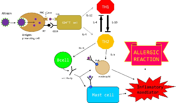
The activated T cell(CD4+ cell) differentiates to Th2 cell by stimulation of interleukin-4(IL-4) called proteins or to Th1 cell by stimulation of IL-12.
Th1 produces IFN-γand IL-2 and lets a macrophage and a killer cell activated and participates in cell-mediated immunity. On the other hand, Th2 produces IL-4, IL-5,IL-10 and lets B cell and a mast cell activated and participates in humoral immunity.
In this way, Th1 and Th2 work important in immunity reaction.
Interaction between Th1 and Th2
By pathogens and stimulation of immunity, a direction of Th1 predominance or the Th2 predominance is decided, and a kind of the immune response is in this way fixed.
When a virus invades the body, differentiation to Th1 is promoted by Il-12 produced by APC and IFN-γ is produced from Th1 and hits infection defense. At the same time, Th2 is controlled by IFN-γ, and the production of cytokine promoting mast cells and eosinophil work is also controlled . On the other hand, when an antigen invades body, differentiation to Th2 is promoted by IL-4 . Th2 produces IL-4, and activate B cells, and B cells produce an antibody (IgE,IgG). Moreover, Th2 produces IL-5 and promotes eosinophil work. At the same time, Th1 is controlled by IL-4, and the production of cytokine promoting the functin of macrophages is also controlled.
In the way, Th1 and Th2 keep balance mutually , and two cells usually control immune response. (Th1/Th2 balance)
When this balance collapses and became the Th2 predominance, IL-5 is secreted excessively from Th2, and eosonophil and mast cells are activated. And than after that inflammatory materials such as histamine or leukotriene are secreted excessively by テthose cells when an antigen is connected in those cells that an antibody bound to through Fc receptor (FcR). This inflammatory material and antibody produced excessively are related to allergic reactions.
The allergic reactions are classified in type Ⅰto Ⅳ ,and not only these reactions occur independently, but also some types may be taking place at the same time.
Above all, type Ⅰ reactions is said immediate hypersensitivity, and time to onset is short.Examples of this allergic reaction include bronchial asthma and atopic dermatitis
There are the people suffering by these allergic diseases.
Therefore, we thought that we kept Th1/Th2 balance normally by increasing IL-12 which participated in Th1 cytodifferentiation and restrained allergic reactions.

Link Light Assay
A variety of protein interaction analysis have been developed.Linklight Assay is a novel method based on an inactive permuted protein which can be activated by a protease to generate stable and sensitive signals for monitoring protein interaction.
Linklight Assay consists of two components: A protein A fused to a Tobacco Etch Virus (TEV) protease and a protein B fused to a permuted luciferase (pLuc) containing a TEV protease cleavage sequence. An inactive pLuc has been created by breaking luciferase into two fragments, rearranging the fragment order in that the N-terminal fragments is moved to C-terminus and C-terminal fragment is moved to the N-terminus and connecting them by a TEV protease cleavage sequence. Upon interaction between protein A and B, inactive pLuc is cleaved, the cleaved luciferase fragments are spontaneously refolded, and active luciferase is reconstituted. Active luciferase works as reporter.
Linklight Assay provides simple, robust, economic, and sensitive methods to detect protein interaction in live cells with immediate and stable signals.
We applied this technology to make IL-5 independent expression system. We will explain our strategy for Ecoli receiving IL5, and producing IL-12 in Strategy Page

HRV protease
TEV protease is a highly sequence-specific cysteine protease from Tobacco Etch Virus (TEV). Due to its high sequence specificity it is frequently used for the controlled cleavage of fusion proteins.
Human Rhinovirus (HRV) 14 3C Protease is a cysteine protease used to remove fusion from proteins with the HRV14 3C cleavage sequence, like TEV protease.HRV14 3C protease is also high specific for the recognition sequence Leu-Glu-Val-Leu-Phe-Gln-↓-Gly-Pro. The 22 kDa protease get its optimal activity at 4 ℃, but also shows a highiy cleavage rate at 37℃.
HRV14 3C protease is very similar to TEV protease. So, we thought that HRV14 3C protease can cleave protease cleavage sequence which connects IL5 receptor and AraC in the same way TEV protease cleavage protein B fused to a pLuc containing a TEV protease cleavage sequence in linklight assay.
Methodology
Strategy for ecoli to receive IL5, and to produces IL-12.
Applying Linklight assay, our goal is to create an ecoli which receives IL-5,and produces IL-12.
IL-5 is an interleukin produced by T helper 2 cells, mast cells and eosinophils. Through binding to the IL-5 receptor, IL-5 stimulates B cells growth and increases immunoglobulin secretion. It is also a key mediator in eosinophils activation.
IL-5receptor consists of a heterodimer of an alpha subunit and a beta subunit. The IL-5 receptor alpha (IL-5Ra) is specific to IL-5. The beta chain (IL-5Rb) is the common beta chain of receptors for IL-3 and granulocyte-macrophage colony-stimulating factor (GM-CSF). After IL-5 binds to IL-5Ra, IL-5Rb meets IL-5 and form heterodimer with alpha subunit. If IL-5 does not bind to IL-5Ra, IL-5Rb cannot form heterodimer with alpha subunit.
The method applied for Ecoli:to receive IL-5and to produce IL-12 is as follows:
Fusing IL-5Ra and IL-5Rb with AraC which contains a HRV protease cleavage sequence and HRV protease respectively. If heterodimer of IL-5Ra and IL-5Rb are formed by IL-5, AraC containing cleavage sequence and HRV are forced into close proximity. HRV protease recognizes cleavage sequence connecting IL5Ra and AraC and cleaves it. AraC is released from IL5Ra by HRV protease. After released from IL5Ra, AraC regulates expression of IL12 coding gene downstream of AraC promoter. When arabinose is present, AraC acts an activator, and activates expression of IL12.HlyA C-terminal signal is a protein secrete tag. So IL12 which has HlyA secrete tag are secreted outside an ecoli. In the lour experiment, we use GFP instead of IL12 to simplify result. This strategy enable for ecoli to receive IL5 and produce IL12.
|