Team:Imperial/Functionalisation
From 2014.igem.org
Team Functionalisation
Overview
Zombie ipsum reversus ab viral inferno, nam rick grimes malum cerebro. De carne lumbering animata corpora quaeritis. Summus brains sit, morbo vel maleficia? De apocalypsi gorger omero undead survivor dictum mauris. Hi mindless mortuis soulless creaturas, imo evil stalking monstra adventus resi dentevil vultus comedat cerebella viventium. Qui animated corpse, cricket bat max brucks terribilem incessu zomby. The voodoo sacerdos flesh eater, suscitat mortuos comedere carnem virus. Zonbi tattered for solum oculi eorum defunctis go lum cerebro. Nescio brains an Undead zombies. Sicut malus putrid voodoo horror. Nigh tofth eliv ingdead.
Key Achievements
Introduction
Bacterial cellulose is already a useful biomaterial as is due its attractive mechanical properties, with research on applications from loudspeaker diaphragms to protective packaging and wound dressings (Lee 2014). However, our project also aims to extend the properties of cellulose by conjugating functional proteins and peptides to it. This allows us to target specific contaminants in water which can not be filtered by size exclusion alone. (refer to water report and informing design sections).
We considered two ways of functionalizing cellulose (figure 1) but continued to work fully on the cellulose-binding domain (CBD) method. The curli-fibre method would be an exciting avenue to explore in the future; it is feasible since there has already been research into making a “programmable” biomaterial from curli fibres using a peptide tag approach (Nguyen et al 2014), and in combination with cellulose could produce a composite with robust mechanical properties as well.

Lorem ipsum dolor sit amet, consectetuer adipiscing elit. Aenean commodo ligula eget dolor. Aenean massa. Cum sociis natoque penatibus et magnis dis parturient montes, nascetur ridiculus mus. Donec quam felis, ultricies nec, pellentesque eu, pretium quis, sem. Nulla consequat massa quis enim. Donec pede justo, fringilla vel, aliquet nec, vulputate eget, arcu. In enim justo, rhoncus ut, imperdiet a, venenatis vitae, justo. Nullam dictum felis eu pede mollis pretium. Integer tincidunt. Cras dapibus. Vivamus elementum semper nisi. Aenean vulputate eleifend tellus. Aenean leo ligula, porttitor eu, consequat vitae, eleifend ac, enim. Aliquam lorem ante, dapibus in, viverra quis, feugiat a, tellus. Phasellus viverra nulla ut metus varius laoreet. Quisque rutrum. Aenean imperdiet. Etia
Aims
Specification
Lorem ipsum dolor sit amet, consectetuer adipiscing elit. Aenean commodo ligula eget dolor. Aenean massa. Cum sociis natoque penatibus et magnis dis parturient montes, nascetur ridiculus mus. Donec quam felis, ultricies nec, pellentesque eu, pretium quis, sem. Nulla consequat massa quis enim. Donec pede justo, fringilla vel, aliquet nec, vulputate eget, arcu. In enim justo, rhoncus ut, imperdiet a, venenatis vitae, justo. Nullam dictum felis eu pede mollis pretium. Integer tincidunt. Cras dapibus. Vivamus elementum semper nisi. Aenean vulputate eleifend tellus. Aenean leo ligula, porttitor eu, consequat vitae, eleifend ac, enim. Aliquam lorem ante, dapibus in, viverra quis, feugiat a, tellus. Phasellus viverra nulla ut metus varius laoreet. Quisque rutrum. Aenean imperdiet. Etia.
Engineering
The cellulose binding domain (CBD) is the link between our functional proteins and the cellulose. Therefore, we wanted to use a CBD which had high affinity and strong binding to cellulose. However, weaker CBDs may still be useful since they could have the potential be controllably eluted under specific conditions, thereby enabling regeneration of the filter when saturated, and controlled disposal of the collected contaminants (refer to water report if relevant?).
We started our search by looking into the parts registry (link to parts.igem.org) and found two CBDs already existed in the registry: CBDclos (link BBa_K863111) and CBDcex (link BBa_K863101). We also added made three new CBDs to contribute to the registry: dCBD (link to below), CBDcipA, and CBDcenA (hyperlink to the relevant sections below). All the CBDs are in RFC(25) freiberg fusion format to allow for ease of use in protein fusions.
CBDclos and CBDcex
These parts were biobricked by Beilefeld 2012 (link to their wiki https://2012.igem.org/Team:Bielefeld-Germany/Results/cbc), please see their part pages (link BBa_K863111 and BBa_K863101) for detailed information. CBDclos is from the cellulolytic bacterium Clostridium cellulovorans and CBDcex originates from the cellulolytic bacterium Cellulomonas fimi. This is the same organism as for CBDcenA; cex is from the exoglucanase gene and cen is from the endoglucanase gene.
Double CBD (dCBD) with N-terminal linker
This part, BBa_K1321340 (link http://parts.igem.org/Part:BBa_K1321340) is based on the double cellulose-binding domain construct synthesised and characterised by Linder et al1996, who found that this double CBD had higher affinity for cellulose than either of the two CBDs on their own. The main difference is that our part contains an additional linker sequence on the N-terminus of the protein.
The two CBDs are from the fungus Trichoderma reesei (Hypocrea jecorina) Exocellobiohydrolase (Exoglucanase) I (cbh1), uniprot ID P62694; and Exocellobiohydrolase (Exoglucanase) II, uniprot ID P07987 (cbh2); with a linker peptide between the two CBDs and at the N-terminus of the protein. Both linkers are the same amino acid sequence and are based on the endogenous linker sequences that exists in cbh1 and cbh2 genes. The linker sequence is PGANPPGTTTTSRPATTTGSSPGP which is the same as used by Linder et al 1996. The first three amino acids are from the cbh2 endogenous linker, and the rest is from the cbh1 endogenous linker. CBDcbh1 is placed C-terminal to CBDcbh2 because naturally CBDcbh1 is a C-terminal domain and CBDcbh2 is an N-terminal domain. Both CBDs are from the CBM family 1 (link to cazy here). The precise location of the CBD within the cbh genes was slightly different according to the uniprot annotations and the sequence used by Linder et al 1996; we chose to use the sequence from the paper since the protein was expressed and characterised successfully.
CBDcpiA with N and C-terminal linker
Description to be completed. An illegal EcoRI site was identified in these parts. A silent mutation can correct this and site-directed mutagenesis is currently in progress with the aim to submit these parts soon.
CBDcenA with C-terminal linker
This CBD is from the Endoglucanase A (cenA) gene from Cellulomonas fimi and also contains an endogenous C-terminal linker. The cellulose binding domain region occurs at the N-terminus of the cenA gene (UniProt P07984 link to http://www.uniprot.org/uniprot/P07984) and is from the CBM family 2 (link to http://www.cazy.org/CBM2.htm)
The linker sequence (PTTSPTPTPTPTTPTPTPTPTPTPTPTVTP) is Pro-Thr box which has an extended conformation and acts as a hinge region (Gilkes et al 1989). The endoglucanase CBDcenA has 50% homology to the exoglucanase CBDcex (BBa_K863101) and the linker is highly conserved in both, but the order of the catalytic, linker and cellulose-binding regions is reversed (Warren et al 1986). It has been shown that CBDcenA has the highest binding affinity for crystalline cellulose out of the C. fimi CBDs (Kim et al 2013).
Functionalisation proteins: Binding partners
Metal Binding Proteins
One of the contaminants we can filter out which current size-exclusion technologies can not target, are heavy metals (link to water report). We have worked with four different metal binding proteins: the metallothioneins SmtA and fMT (link to below), a histidine-rich nickel binding protein (NiBP) (link to below) from the bacterium Heliobacter pylori and a synthetic phytochelatin (PC) EC20 (link to below).
SmtA and fMT metallothioneins
These two parts already existed in the BioBrick registry, but since we needed to use them as fusions with the CBDs we improved the parts by making them compatible with the RFC25 (link) Frieburg Fusion BioBrick standard.
Metallothioneins (MTs) are genetically encoded cysteine-rich peptides which serve a role in the detoxification of heavy metals in plants, animals and some prokaryotes (Cobbett, 2002). They have a similar structure and function to phytochelatins, but are produced by different mechanisms (Cobbett, 2002). SmtA is from the cyanobacteria Synechococcus sp. and was used by the Tokyo-NoKoGen 2011 igem team (link) (who submitted the RFC10 part originally, BBa_K519010) to confer cadmium (Cd2+) tolerance to E.coli cells when expressed. fMT originates from the seaweed Fucus vesiculosus and was submitted by Groningen team 2009 (link) and detailed information can be found on the original part page (BBa_K190019).
We successfully cloned the following CBD fusion constructs: (insert table when finalised)
Nickel binding protein (NiBP)
This protein is the histidine-rich nickel-binding protein from Heliobacter pylori (GenBank accession ACX98466.1), and already existed as a BioBrick in the registry (link to K1151001). Since we needed to use this part in fusion proteins we improved the part by submitting it as a basic part in RFC(25) (link) Freiberg fusion format (link here to K1321009).
We successfully cloned (link to cloning protocols on the word cloned) the following CBD fusion constructs with NiBP: (insert table from the parts table spreadsheet when finalised)
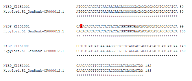
However, sadly we discovered late on in the project that the DNA sequence of the NiBP protein in the registry is out of frame, leading to an incorrect protein translation and read-through of the stop codon (figure 2).

Further investigation into the error let us to identify a single nucleotide deletion in BBa_K1151001 compared to the native H.pylori DNA sequence (GenBank CP000012.1 (link http://www.ncbi.nlm.nih.gov/nuccore/261837457) bp1391851-1392033, reverse complement strand). As seen in Figure 3 the deletion is a ‘C’ at bp 53 of the native sequence. This error was propagated through all our NiBP constructs and means that any subsequent C-terminal fusions are also out of frame and we were unable to express and characterise these constructs. We have noted this on the description pages for each of the parts and hope that in the future they will still be useful, since the error could be corrected relatively easily by a team who wishes to use them by site-directed-mutagenesis PCR to insert the missing nucleotide.
IMAGE right aligned 50% width
Synthetic phytochelatin EC20
This is our new contribution of a metal-binding protein to the parts registry. Synthetic phytochelatins are analogues of phytochelatins (PCs); cysteine-rich peptides of the general structure (γ-GluCys)n-Gly (n~2-5) which bind and sequester heavy metals, with a role in detoxification (Cobbett & Goldsbrough 2002). PCs are found mainly in plants but also in other organisms including diatoms, fungi, algae (including cyanobacteria) and some invertebrates (Cobbett & Goldsbrough 2002, Rea 2012). Natively, the PCs are enzymatically synthesized by phytochelatin synthase (PCS) from a glutathione (GSH) precursor and the amino acids in the resulting peptide are linked by the non-standard gamma peptide bonds (Cobbett & Goldsbrough 2002, Rea 2012). However, genetically encoded analogues (containing normal alpha-peptide bonds) of different number Glu-Cys (EC) repeats have been characterised, with the EC20 having the highest binding (Bae et al 2000).
The main metal PCs confer tolerance to is Cadmium (Cobbett & Goldsbrough 2002) with a reported stoichiometry of 10 Cd2+ per peptide (Bae et al, . EC20 peptide has still showed good metal ion binding whilst fused to a cellulose binding domain (CBD) for water purification (Xu et al 2002) and when anchored to bacterial cell membrane to confer Cd2+ tolerance (Bae et al 2000, Chaturvedi & Archana 2014). PC EC20 has also been shown to bind many more heavy metals. For example, when displayed on the surface of Cupriavidus metallidurans, the cells capacity to adsorb ions increased by 31 to 219% depending on the ion (table 1, Biondo et al 2012). These binding properties of PC EC20 have been utilised in applications including a regeneratable biosensor of Hg2+, Cd2+, Pb2+, Cu2+ and Zn2+ ions to 100 fM–10 mM (Bontidean et al 2003), and to target CdSe/ZnS quantum dots to streptavidin expressing HeLa cells when biotinylated (Pinaud et al 2004). INSERT TABLE OF THE IONS FROM THE PAPER
We used this part to create a library of different cellulose-binding domains fused to phytochelatin: (insert parts table from the spreadsheet here when it is finalised)
Other potential proteins
During our research we also explored other functional proteins to fuse to the CBDs.
Laccases
Laccases are enzymes which can break down aromatic and phenolic compounds such as modified estrogens which can end up unfiltered in the water supply with unknown consequences (Auriol et al 2008). This method of treating estrogen in water was explored by Bielefeld 2012 iGEM team (link), and our system also has potential to address this.
Nanobodies
Nanobodies are fragments of camelid antibodies which can be generated against a variety of antigens and can be easily expressed by organisms such as E. coli . Nanobodies could be used to target viruses, which are often too small to be filtered by size exclusion. For example there are already nanobodies which can bind to Vaccinia virus, ricin, Cholera toxin, Stephylococcal enterotoxin B and H5N1 Influenza (Goldman et al 2006, Ibanez et al 2011) to name a few. Additionally, nanobodies are stable and can be selected to resist proteolytic attach (Harmsen et al 2006). Nanobodies can also be functional when as fusions or conjugates to other proteins for example to target enzymes to specific cells (Cortez-Retamozo 2004), so there is much potential for targeting a wide array of contaminants when fused to cellulose binding domains.
Modelling
Lorem ipsum dolor sit amet, consectetuer adipiscing elit. Aenean commodo ligula eget dolor. Aenean massa. Cum sociis natoque penatibus et magnis dis parturient montes, nascetur ridiculus mus. Donec quam felis, ultricies nec, pellentesque eu, pretium quis, sem. Nulla consequat massa quis enim. Donec pede justo, fringilla vel, aliquet nec, vulputate eget, arcu. In enim justo, rhoncus ut, imperdiet a, venenatis vitae, justo. Nullam dictum felis eu pede mollis pretium. Integer tincidunt. Cras dapibus. Vivamus elementum semper nisi. Aenean vulputate eleifend tellus. Aenean leo ligula, porttitor eu, consequat vitae, eleifend ac, enim. Aliquam lorem ante, dapibus in, viverra quis, feugiat a, tellus. Phasellus viverra nulla ut metus varius laoreet. Quisque rutrum. Aenean imperdiet. Etia
Testing
Lorem ipsum dolor sit amet, consectetuer adipiscing elit. Aenean commodo ligula eget dolor. Aenean massa. Cum sociis natoque penatibus et magnis dis parturient montes, nascetur ridiculus mus. Donec quam felis, ultricies nec, pellentesque eu, pretium quis, sem. Nulla consequat massa quis enim. Donec pede justo, fringilla vel, aliquet nec, vulputate eget, arcu. In enim justo, rhoncus ut, imperdiet a, venenatis vitae, justo. Nullam dictum felis eu pede mollis pretium. Integer tincidunt. Cras dapibus. Vivamus elementum semper nisi. Aenean vulputate eleifend tellus. Aenean leo ligula, porttitor eu, consequat vitae, eleifend ac, enim.
Results
Lorem ipsum dolor sit amet, consectetuer adipiscing elit. Aenean commodo ligula eget dolor. Aenean massa. Cum sociis natoque penatibus et magnis dis parturient montes, nascetur ridiculus mus. Donec quam felis, ultricies nec, pellentesque eu, pretium quis, sem. Nulla consequat massa quis enim. Donec pede justo, fringilla vel, aliquet nec, vulputate eget, arcu. In enim justo, rhoncus ut, imperdiet a, venenatis vitae, justo. Nullam dictum felis eu pede mollis pretium. Integer tincidunt. Cras dapibus. Vivamus elementum semper nisi. Aenean vulputate eleifend tellus. Aenean leo ligula, porttitor eu, consequat vitae, eleifend ac, enim.
References
Lee, K.-Y., Buldum, G., Mantalaris, A. and Bismarck, A. (2014), More Than Meets the Eye in Bacterial Cellulose: Biosynthesis, Bioprocessing, and Applications in Advanced Fiber Composites. Macromol. Biosci., 14: 10–32. doi: 10.1002/mabi.201300298 Nguyen, P.Q., Botyanszki, Z., Tay, P.K.R. & Joshi, N.S. (2014) Programmable biofilm-based materials from engineered curli nanofibres. Nature communications. [Online] 54945. Available from: doi:10.1038/ncomms5945. Zakeri, B., Fierer, J.O., Celik, E., Chittock, E.C., et al. (2012) Peptide tag forming a rapid covalent bond to a protein, through engineering a bacterial adhesin. Proceedings of the National Academy of Sciences of the United States of America. [Online] 109 (12), E690–E697. Available from: doi:10.1073/pnas.1115485109. Linder, M.; Salovuori, I.; Ruohonen, L.; Teeri, T.T., 1996. Characterization of a Double Cellulose-binding Domain. SYNERGISTIC HIGH AFFINITY BINDING TO CRYSTALLINE CELLULOSE. Journal of Biological Chemistry, 271(35), pp.21268–21272. Available at: http://www.jbc.org/content/271/35/21268.full Gilkes, N.R., Kilburn, D.G., Miller, R.C. & Warren, R.A. (1989) Structural and functional analysis of a bacterial cellulase by proteolysis. The Journal of biological chemistry. 264 (30), 17802–17808. Warren, R.A., Beck, C.F., Gilkes, N.R., Kilburn, D.G., et al. (1986) Sequence conservation and region shuffling in an endoglucanase and an exoglucanase from Cellulomonas fimi. Proteins. 1 (4), 335–341. Kim, H.-D., Choi, S.-L., Kim, H., Sohn, J.H., et al. (2013) Enzyme-linked assay of cellulose-binding domain functions from Cellulomonas fimi on multi-well microtiter plate. Biotechnology and Bioprocess Engineering. 18 (3), 575–580. Cobbett, C. & Goldsbrough, P. (2002) Phytochelatins and metallothioneins: roles in heavy metal detoxification and homeostasis. Annual review of plant biology. [Online] 53159–182. Available from: doi:10.1146/annurev.arplant.53.100301.135154. Rea, P.A. (2012) Phytochelatin synthase: of a protease a peptide polymerase made. Physiologia plantarum. 145 (1), 154–164. Bae, W., Chen, W., Mulchandani, A. & Mehra, R.K. (2000) Enhanced bioaccumulation of heavy metals by bacterial cells displaying synthetic phytochelatins. Biotechnology and bioengineering. 70 (5), 518–524. Xu, Z., Bae, W., Mulchandani, A., Mehra, R.K., et al. (2002) Heavy Metal Removal by Novel CBD-EC20 Sorbents Immobilized on Cellulose. Biomacromolecules. [Online] 3 (3), 462–465. Chaturvedi, R. & Archana, G. (2014) Cytosolic expression of synthetic phytochelatin and bacterial metallothionein genes in Deinococcus radiodurans R1 for enhanced tolerance and bioaccumulation of cadmium. Biometals : an international journal on the role of metal ions in biology, biochemistry, and medicine. 27 (3), 471–482. Biondo, R., da Silva, F.A., Vicente, E.J., Souza Sarkis, J.E., et al. (2012) Synthetic phytochelatin surface display in Cupriavidus metallidurans CH34 for enhanced metals bioremediation. Environmental science & technology. 46 (15), 8325–8332. Bontidean, I., Ahlqvist, J., Mulchandani, A., Chen, W., et al. (2003) Novel synthetic phytochelatin-based capacitive biosensor for heavy metal ion detection. Biosensors and Bioelectronics. 18 (5-6), 547–553. Pinaud, F., King, D., Moore, H.-P. & Weiss, S. (2004) Bioactivation and cell targeting of semiconductor CdSe/ZnS nanocrystals with phytochelatin-related peptides. Journal of the American Chemical Society. 126 (19), 6115–6123. Auriol, M., Filali-Meknassi, Y., Adams, C.D., Tyagi, R.D., et al. (2008) Removal of estrogenic activity of natural and synthetic hormones from a municipal wastewater: efficiency of horseradish peroxidase and laccase from Trametes versicolor. Chemosphere. [Online] 70 (3), 445–452. Available from: doi:10.1016/j.chemosphere.2007.06.064. Harmsen, M.M., van Solt, C.B., van Zijderveld-van Bemmel, A.M., Niewold, T.A., et al. (2006) Selection and optimization of proteolytically stable llama single-domain antibody fragments for oral immunotherapy. Applied microbiology and biotechnology. [Online] 72 (3), 544–551. Available from: doi:10.1007/s00253-005-0300-7. Cortez-Retamozo, V. (2004) Efficient Cancer Therapy with a Nanobody-Based Conjugate. Cancer Research. [Online] 64 (8), 2853–2857. Available from: doi:10.1158/0008-5472.CAN-03-3935.
 "
"


