Team:Paris Saclay/Protocols/Gel Electrophoresis
From 2014.igem.org
Gel Electrophoresis
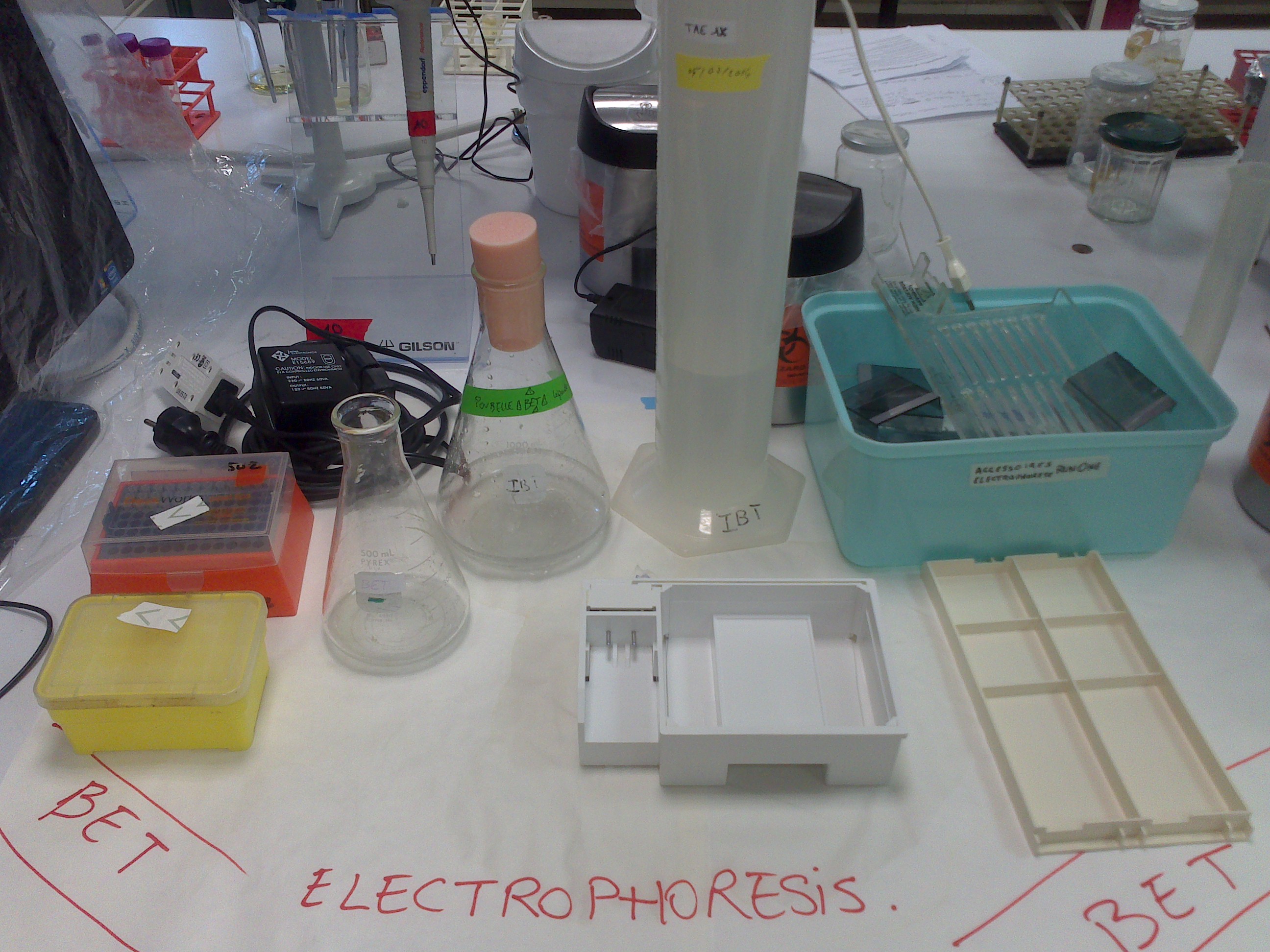
Gel electrophoresis is a widely used technique for the analysis of nucleic acids and proteins. Almost every molecular biology research laboratory routinely uses agarose gel electrophoresis for the purification and analysis of DNA. We will be using agarose gel electrophoresis to determine the presence, the quantity and size of PCR products and digested DNA fragments.
- Preparing 1% agarose gel:
- Weight out 1g of Agarose powder and add it to a 500 ml flask.
- Add 100 ml of TAE Buffer to the flask.
- Melt the agarose in a microwave until the solution become clear (heat the solution for several short intervals - do not let the solution boil for long periods as it may boil out of the flask).
- Let the solution cool to about 50-55°C, swirling the flask occasionally to cool it evenly.
- Add BET with a final concentration of 200ng/µL.
- Place the combs in the gel casting tray.
- Pour the melted agarose solution into the casting tray and let cool until it is solid (it should appear milky white).
- Carefully pull out the combs and remove the tape.
- Loading Samples and Running an Agarose Gel:
- Place the gel in the electrophoresis chamber.
- Add enough TAE Buffer so that there is about 2-3 mm of buffer over the gel.
- Add loading buffer 6X to each of your samples.
- Carefully load a molecular weight ladder into the first lane of the gel.
- Carefully load your samples into the additional wells of the gel.
- Record the order each sample will be loaded on the gel, including who prepared the sample, the DNA template - what organism the DNA came from, controls and ladder
- Run the gel usually at 100V during 30 mins or until the dye line is approximately 75-80% of the way down the gel.
- Turn OFF power, disconnect the electrodes from the power source, and then carefully remove the gel from the gel box.
- Using any device that has UV light, visualize your DNA fragments.
 "
"


