Team:Marburg:Project:Silver
From 2014.igem.org
Silver Surfer
"Surfing is a surface water sport in which the wave rider, referred to as a surfer, rides on the forward or deep face of a moving wave, which is usually carrying the surfer towards the shore."
In modern science at the limits of visualization and detection feasibility the
amplification of signals is one of the challenges. Scaffolding – building a high
density of factors in close proximity – is a clue to optimize chemical reactions or
increasing signals for detection. Here we use a natural structure: the bacterial
flagellum as a toolbox for different applications. The bacterial flagellum consists
of the basal body, the hook and the filament – at which the filament is built out
of 20.000 copies of Flagellin. Due to the rotating flagellum bacteria are able to
surf through the environment (Fig1).
 Figure 1: Composition of the bacterial Flagellin filament.
Among Bacteria Flagellin is a highly conserved protein and consists of four domains (D0-D3) in which only the D2 and D3 domain are variable, visualized in Figure 2 (Yonekura et al. 2003). By comparison of Salmonella, Sphingomoas and Bacillus Flagellin it is remarkably that Bacillus D2 and D3 is reduced to a small loop (Fig. 2) (Altegoer et al. 2014).
 Figure 2: Conserved protein domains of Flagellin among different bacteria.
This fact was the reason to have a closer look at the Flagellin of Bacillus subtilis. We have the vision of using the bacterial flagellum as a scaffold for high density accumulation of high value application proteins.
“SURFing (Synthetic Units for Redirecting Functionalities) is the redirection of the natural flagellum structure to a high density surface which can be modified for different purposes, forward to solve important challenges of the humanity e.g. in reduction of environmental pollution and health applications. (Fig. 3)” (iGEM Marburg 2014).
 Figure 3: SURFing application workflow.
As a proof of principle we forced ourselves to contribute to the reduction of environmental pollution regarding heavy metal ions. Silver ions have a high toxicity and are lethal in high dose. This effect is on the one hand industrially welcome and used as additives on the other hand it is a big problem due to industrial wastewater. This is the reason why the SilverSURF project was born.
SilverSURF
The aim of the SilverSURF project is to catch ions from soil and solutions and bind them with a high affinity. How should this be realized? Our vision is to insert an additional modified Flagellin copy into the genome of Bacillus subtilis. This allele should be under control of a silver ion sensitive promoter. By exposure to silver ions the modified Flagellin is expressed and incorporated into the filament. This modified Flagellin is a chimeric protein with a new domain – the metallothionein Cup1 from yeast which specifically binds silver and copper ions (Moore et al. 2005). This leads to engineered B. subtilis cells which catch the surrounding ions (Fig. 4).

 Figure 4: SilverSURF vision.
Although there is published data about specific transcriptional changes due to heavy metal ion stress in B. subtilis we were not able to find a specific promoter. We established a reporter system to analyze different promoters, the promoter of the argJ for silver and the promoter of the yknO for copper, by generating a highly modular amyE locus integration plasmid with a GFP reporter. By analyzing our reporter constructs via STED microscopy we could show that the chosen silver sensitive promoter does not show any differences by exposure to different silver concentrations compared to our negative control without silver ions (Fig 5). In principle the established promoter reporter system works though.
 Figure 5: SilverSURF promoter reporter system with silver concentrations 0, 0.1 µM and 10 µM.
Nevertheless we focused onto the Flagellin Cup1 chimera (Moore et al. 2005). We designed a modular system for chromosomal integration where the hag gene which codes for Flagellin features a Spe1 site for fast and modular exchange of domains via Gibson Assembly (pIGEM-0016). The Cup1 module was amplified from Saccharomyces cerevisiae genomic DNA integrated into the pIGEM-0016 system and the wild type hag successfully replaced with the chimeric hag-cup construct. pMAD allows highly efficient allelic replacement of the wild type hag via the pMAD system (Arnaud et al. 2004).
Motility Assays and structural analysis
To test whether the engineered Flagellin is functional in vivo swimming and swarming assays were performed. Unfortunately the assays showed that the engineered Flagellin containing strains were strongly reduced in their motility compared to both the wild type strain WT3610 and the control strain producing our modularised Flagellin Hag-Spe1 without the Cup1 domain.
Anyway, to get a clue what might disrupt the Flagellin functionality the Hag-Cup1 was overexpressed in Escherichia coli. Sufficient amounts of protein could be purified which were subjected to crystallization to solve the structure of the chimeric protein. Crystals could be obtained but the structure could not be solved due to the disruption of D1 domain by the Cup1 integration into Hag.
This results leads us to rethink our approach and we concluded that it might work if we replace the Bacillus loop with the D2 Domain of Salmonella typhimurium (Fig. 2) and use this as a linker for the Cup1 (Fig. 6). This construct is further on called Hag-D2-Cup1.
 Figure 6: New engineered synthetic D2-Cup1 Flagellin.
At the same time we had the idea to make a Hag-D2-Strep construct which would give us flexibility by coupling high value applicable proteins to streptavidin and further on build an extracellular complex of Flagellin with chimeric streptavidin protein (for further information see the CancerSURF project).
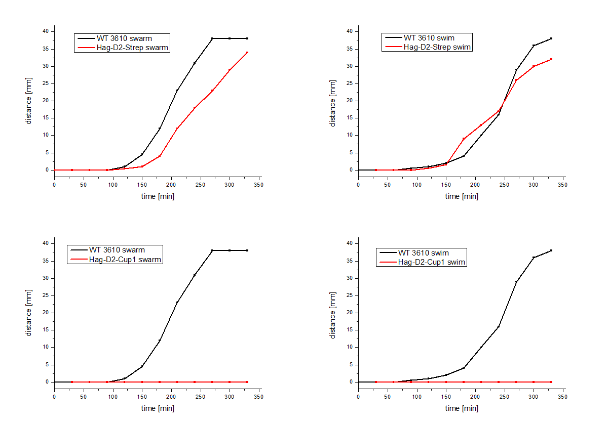
The Hag-D2-Cup1 and Hag-D2-Strep were integrated into B. subtilis via homologous recombination by using again our modular pMAD system. Again motility assays were performed and these show similar results for Hag-Cup1 and Hag-D2-Cup1 though to our surprise the Hag-D2-Strep is wild type like in this assay.
 Figure 7: Motility assays of the Hag-D2-Cup1 and the Hag-D2-Strep.
This leads us to the question why does one construct show a wild type like phenotype but the other not. Therefore, we visualized the flagella by using uranylacetate negative stained electron microscopy (Figure 7). To our surprise we could show that the Hag-D2-Cup1 strain is able to build filaments though they look different to the wild type. They are much shorter and inflexible which might be the reason why we observed fragmented filaments. Astonishingly the Hag-D2-Strep filaments look like wild type filaments. Although we observed less filaments they seem to be functional. What might be the difference between Hag-D2-Cup1 and Hag-D2-Strep is part of further investigations.
 Figure 8: Uranylacetate negative stained electron microscopy of B. subtilis (A) and engineered strains (B).
The successful engineering of the Hag-D2-Strep Flagellin is the corner stone for the success story of our CancerSURF project. In further investigations we plan to couple Cup1-Streptavidin to harvested Hag-D2-Strep f ilaments for our SilverSURF – this would directly optimize our approach for pollution reduction. Beyond that there is no need to use the GMO outside the laboratory. The recombinant proteins could be used directly so that there are no emerging safety concerns. Also the higher level of modularity allows us to couple every imaginable high value applicable protein to our filaments. There is no reason to use only one protein, we also plan to use different proteins coupled to streptavidin and bring them into close proximity by using the filaments as a scaffold for molecular crowding. There are unlimited high valuable applications for such a system to solve all kinds of challenges the humanity is faced with.
Altegoer, Florian; Schuhmacher, Jan; Pausch, Patrick; Bange, Gert (2014): From molecular evolution to biobricks and synthetic modules: a lesson by the bacterial flagellum. In: Biotechnology and Genetic Engineering Reviews 30 (1), S. 49–64. DOI: 10.1080/02648725.2014.921500.
Arnaud, Maryvonne; Chastanet, Arnaud; Débarbouillé, Michel (2004): New vector for efficient allelic replacement in naturally nontransformable, low-GC-content, gram-positive bacteria. In: Appl. Environ. Microbiol. 70 (11), S. 6887–6891. DOI: 10.1128/AEM.70.11.6887-6891.2004.
Moore, Charles M.; Gaballa, Ahmed; Hui, Monica; Ye, Rick W.; Helmann, John D. (2005): Genetic and physiological responses of Bacillus subtilis to metal ion stress. In: Molecular Microbiology 57 (1), S. 27–40, zuletzt geprüft am 16.10.2014.
Yonekura, Koji; Maki-Yonekura, Saori; Namba, Keiichi (2003): Complete atomic model of the bacterial flagellar filament by electron cryomicroscopy. In: Nature 424 (6949), S. 643–650. DOI: 10.1038/nature01830.
 "
"