Team:Austin Texas/kit
From 2014.igem.org
| Line 164: | Line 164: | ||
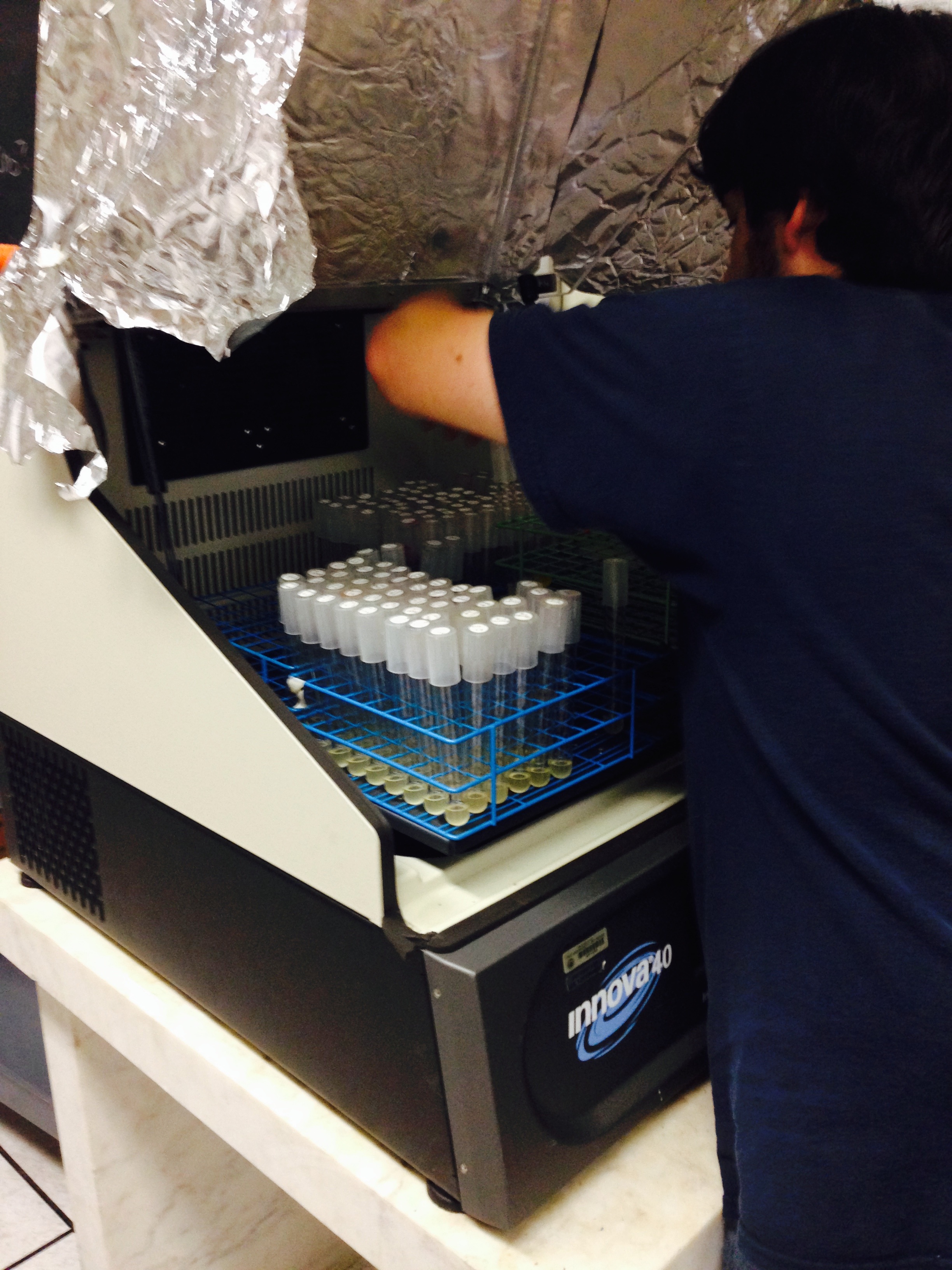
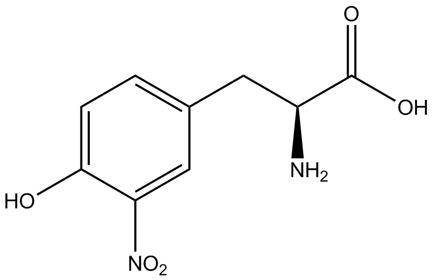
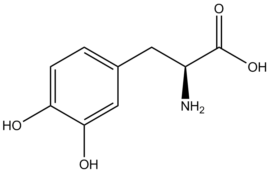
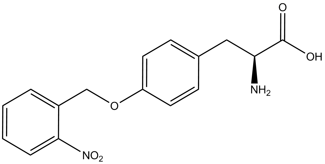
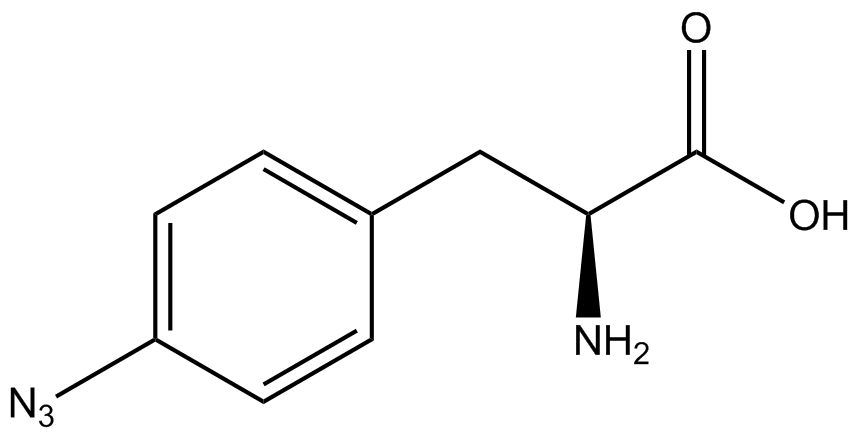
ncAA-specific concerns accounted for and should be noted. These include: light-sensitivity, oxidation, and interference with fluorescence readings. Certain ncAAs were protected from light due to their light-sensitive molecular structures such as [https://2014.igem.org/Team:Austin_Texas/kit#ncAA_Table ONBY and AzF]. These ncAAs were prepared in a dark room and wrapped in foil. All cultures were grown in a foil-wrapped incubator for consistency. Some ncAAs are more prone to oxidation, specifically L-dopa. When oxidized, the solution turns black. To prevent oxidation, each ncAA was prepared the day of the test for consistency. By this method, the oxidation of L-dopa did not occur quickly enough to have an effect on the data. Most ncAA solutions were transparent once prepared with the exception of 3-nitrotyrosine. The yellow-orange tint of 3-nitrotyrosine solution was accounted for by measuring the fluorescence of 1mM 3-nitrotyrosine in media and subtracting any possible background fluorescence from culture fluorescence grown in 1mM 3-nitrotyrosine. | ncAA-specific concerns accounted for and should be noted. These include: light-sensitivity, oxidation, and interference with fluorescence readings. Certain ncAAs were protected from light due to their light-sensitive molecular structures such as [https://2014.igem.org/Team:Austin_Texas/kit#ncAA_Table ONBY and AzF]. These ncAAs were prepared in a dark room and wrapped in foil. All cultures were grown in a foil-wrapped incubator for consistency. Some ncAAs are more prone to oxidation, specifically L-dopa. When oxidized, the solution turns black. To prevent oxidation, each ncAA was prepared the day of the test for consistency. By this method, the oxidation of L-dopa did not occur quickly enough to have an effect on the data. Most ncAA solutions were transparent once prepared with the exception of 3-nitrotyrosine. The yellow-orange tint of 3-nitrotyrosine solution was accounted for by measuring the fluorescence of 1mM 3-nitrotyrosine in media and subtracting any possible background fluorescence from culture fluorescence grown in 1mM 3-nitrotyrosine. | ||
| + | |||
RFP fluorescence was measured using: excitation - 550 nm, emission - 675 | RFP fluorescence was measured using: excitation - 550 nm, emission - 675 | ||
| + | |||
GFP fluorescence was measured using: excitation - 480 nm, emission - 525 | GFP fluorescence was measured using: excitation - 480 nm, emission - 525 | ||
Revision as of 17:05, 17 October 2014
| |||||||||||||||||||||||||||||||||||||||||||||||||||||||||||||||||||||||||||||||||||||||||||||||||||||
 "
"

