Team:Austin Texas/photocage
From 2014.igem.org
Nathanshin (Talk | contribs) |
|||
| Line 106: | Line 106: | ||
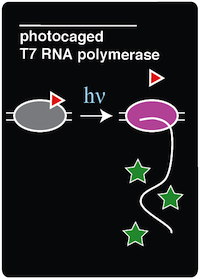
In order to create an in vivo light-activated GFP expression system, two plasmids were constructed and transformed into amberless E.coli cells. The first plasmid contains the tRNA and synthetase pair, which is necessary to incorporate ONBY into the amber stop codon of the T7 RNAP. The second plasmid contains the coding sequence for T7 RNAP, which has a mutation on the Tyrosine 639 residue, and a GFP coding sequence bound to an upstream T7 promoter. Once these components were assembled by Gibson Assembly, the two plasmids were transformed via electroporation into aliquots of Amberless E.coli. | In order to create an in vivo light-activated GFP expression system, two plasmids were constructed and transformed into amberless E.coli cells. The first plasmid contains the tRNA and synthetase pair, which is necessary to incorporate ONBY into the amber stop codon of the T7 RNAP. The second plasmid contains the coding sequence for T7 RNAP, which has a mutation on the Tyrosine 639 residue, and a GFP coding sequence bound to an upstream T7 promoter. Once these components were assembled by Gibson Assembly, the two plasmids were transformed via electroporation into aliquots of Amberless E.coli. | ||
| + | For this experiment, there were also other necessary control strains to test alongside the experimental strain. These controls included a T7-GFP construct with no amber stop codon in the O-helix (to serve as a positive control for expression with T7 polymerase), sfGFP amberless E.coli (to observe the expression of GFP by native polymerase), amberless E.coli (to serve as a cell background control), and LB supplemented with ncAA (to serve as a media background control). | ||
| - | + | All of these strains were grown overnight in LB and appropriate antibiotics. The next morning, 100 microliters of each of the strains were pipetted into three different conditions. The conditions are as follows: | |
| - | + | *(-)IPTG, (-)ONBY | |
| - | + | *(+)IPTG, (+)ONBY | |
| - | + | *(+)IPTG, (+) ONBY | |
| + | Each of the conditions were in sterile test tubes and contained 1 mL of media and appropriate antibiotics. These cultures were allowed to grow for 2 hours. After 2 hours of growth, IPTG was added to all (+)IPTG condition tubes. The test tubes were allowed to grow another 2-4 hours, or until the OD600 reached 0.3. At this point, five 100 microliter samples of each of the strains were transferred into a 96-well plate. | ||
After irradiation with light, these cells were allowed to grow overnight before taking fluorescent measurements. This additional growth was necessary to allow the "decaged" T7 RNAP to polymerize mRNA transcripts of the GFP coding sequence. | After irradiation with light, these cells were allowed to grow overnight before taking fluorescent measurements. This additional growth was necessary to allow the "decaged" T7 RNAP to polymerize mRNA transcripts of the GFP coding sequence. | ||
Revision as of 03:15, 17 October 2014
| |||||||||||||||||||||||||||||
 "
"


