should have gel image of OmpA-Bovine as well


A B
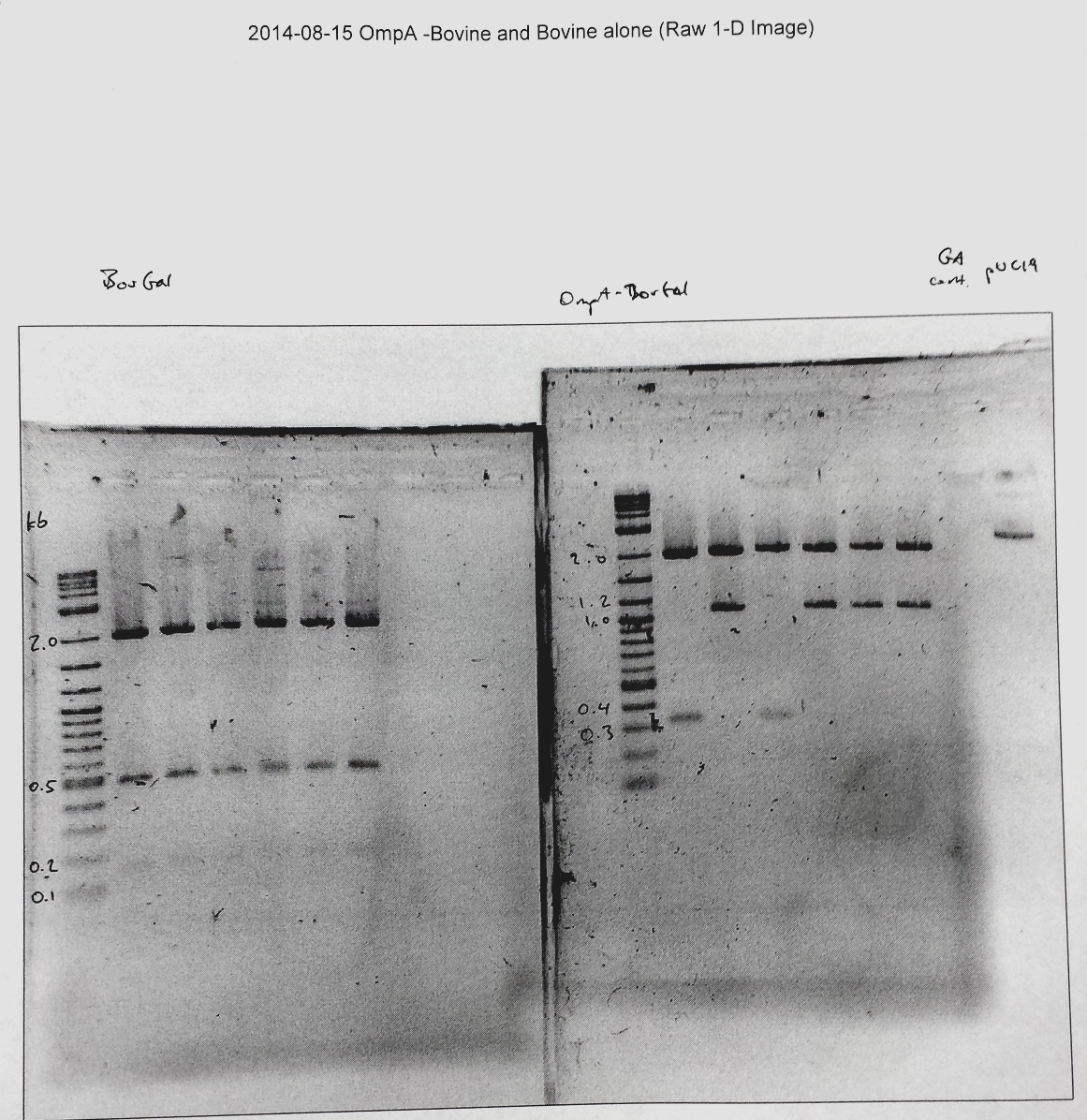
Fig. 1 - Bovine Galectin-1 gene cloned into the pSB1C3 vector with pBAD promoter and a RBS
Galectin-1 from Bos taurus has been cloned into pSB1C3 vector. Bos taurus or bovine galectin enabled us to generate a platform that enables future work on ligand binding. While CvGals binding characteristics are still being researched the crystal structure and nature of bovine galectin has been well studied. The subsequent BioBricks regarding this gene were all constructed via Gibson assembly and cloning. The plasmids from which the OmpA insert and the vector were amplified via extension PCR are respectively: OmpA with linker (BBa_K103006) and pBAD-RBS on the pSB1C3 backbone (BBa_K750000). For the latter, the GFP in the BioBrick was not included for Gibson assembly. The bovine galectin-1 was ordered as a synthetically synthesized sequence from IDT as a linear gene and amplified directly with appropriate primers for Gibson assembly.
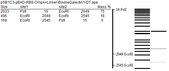
Figure 1A shows the Bovine galectin-1 assembled into pSB1C3-pBAD-RBS alone (no OmpA), which when expressed in E. coli should produce a soluble form of the protein. After successful assembly and cloned into DH5α E. coli. The plasmid was cut with restriction enzymes EcoRI and PstI, standard sites in the pSB1C3 vector’s BioBrick prefix and suffix, respectively. The result is expected to yield the insert, pBAD-RBS-Bovine galectin-1, and the pSB1C3 vector. However, an unwanted EcoRI site was found in the galectin-1 gene afterwards, yielding the pattern seen in the gel. Figure 1B demonstrates the expected pattern, with band sizes of 2033 bp, 496 bp, and 169 bp top to bottom, via the software “A plasmid Editor” (ApE, courtesy of M. Wayne Davis, University of Utah).
 B
B
Fig. 2 - need gel of OmpA-Bovine (cut with EcoRI and PstI, before SDM)
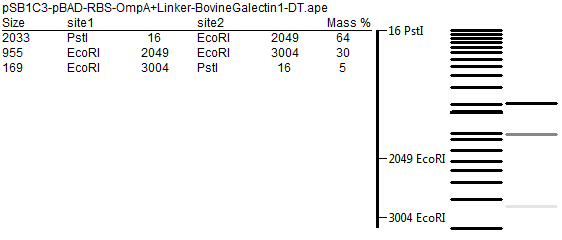
Figure 2A shows the Bovine galectin-1 assembled into pSB1C3-pBAD-RBS as a fusion protein with OmpA. When expressed in E. coli, the galectin should continue off the C-terminus of the OmpA-linker and thus be transported to the extracellular matrix since it is attached to the OmpA transport protein. After plasmid assembly transfer of plasmid into a DH5α E. coli cells strain, the plasmid was extracted and digested with EcoRI and PstI. The pattern of three bands appears again due to an additional EcoRI restriction site.
Fig. 3 - sequencing result/chromatogram of SDM’d plasmids? or BLAST result (BBa_K1489000 and BBa_K1489004)
To address this issue, site-directed mutagenesis was performed on these plasmids to change the EcoRI sequence within the bovine galectin-1 gene (5’-GAATTC-3’) to 5’-GAGTTT-3’, which will code for the same Glu-Phe sequence. Figure 3A and 3B demonstrate the two successfully mutagenized base pairs in the bovine galectin-1 gene, while preserving the rest of the plasmid.
Fig 4. - sequence/chromatogram of OmpA moved into pSB1C3
Since UMaryland worked extensively with the OmpA-linker (BBa_K103006), we have also looked into improvements on that BioBrick.
Fig. 5 - sequence/chromatogram of SDM’d OmpA in pSB1C3
 "
"