Team:SUSTC-Shenzhen/Notebook/HeLaCell/Construct-Cellline
From 2014.igem.org
(minor add) |
|||
| Line 23: | Line 23: | ||
:''Procedure'' | :''Procedure'' | ||
# Wash cells with PBS, trypsin digestion, and medium to stop digestion. | # Wash cells with PBS, trypsin digestion, and medium to stop digestion. | ||
| - | # Transfer the sell suspension in 15ml centrifuge tube and centrifuge at 800rpm, | + | # Transfer the sell suspension in 15ml centrifuge tube and centrifuge at 800rpm, for 5 min. |
# Discard the supernatant and add 2ml complete medium to re suspend the cells. | # Discard the supernatant and add 2ml complete medium to re suspend the cells. | ||
# Add 1ml PBS to every gap between well | # Add 1ml PBS to every gap between well | ||
| Line 69: | Line 69: | ||
# Cas9(Cas9+TetOn) & Piggybac | # Cas9(Cas9+TetOn) & Piggybac | ||
For the first two combination we have different ratio of EGFP to get mono-copy of EGFP in cell line. | For the first two combination we have different ratio of EGFP to get mono-copy of EGFP in cell line. | ||
| + | |||
<html><div class="table-responsive"></html> | <html><div class="table-responsive"></html> | ||
{|class="table" | {|class="table" | ||
| Line 150: | Line 151: | ||
[2014 Aug 16th 9:00am] | [2014 Aug 16th 9:00am] | ||
| - | '''''The | + | '''''The first day after transfection''''' |
Change the medium of transfected cells and observe the fluorescent with fluorescent microscope. | Change the medium of transfected cells and observe the fluorescent with fluorescent microscope. | ||
| Line 157: | Line 158: | ||
The transfected cells are observed before we change the medium. | The transfected cells are observed before we change the medium. | ||
| - | |||
| + | {SUSTC-Image|wiki/images/0/05/SUSTC-Shenzhen-3DayAfterTransefation-8.17G1.tif|G1 3day} | ||
| + | Figure 1. | ||
| + | //TODO figure | ||
| Line 167: | Line 170: | ||
[2014 Aug 16th 15:00pm] | [2014 Aug 16th 15:00pm] | ||
| - | '''''The | + | '''''The first day after transfection''''' |
Passage part of cells to the 10cm petri dish (for picking monoclonal later except G5), and the remaining cells are still seeded on the previous 24-well plates (about 500μL cell suspension per well). (15:00 pm – 18:00 pm) | Passage part of cells to the 10cm petri dish (for picking monoclonal later except G5), and the remaining cells are still seeded on the previous 24-well plates (about 500μL cell suspension per well). (15:00 pm – 18:00 pm) | ||
| Line 217: | Line 220: | ||
[2014 Aug 18th 10:00am] | [2014 Aug 18th 10:00am] | ||
| - | '''''The | + | '''''The third day after transfection''''' |
Observe the transfected cells on the plate under fluorescence microscope. | Observe the transfected cells on the plate under fluorescence microscope. | ||
| - | // | + | |
| + | {{SUSTC-Image|wiki/images/0/05/SUSTC-Shenzhen-3DayAfterTransefation-8.17G1.tif|G1 3days}} | ||
| + | Figure . The fluorescent picture of G1 captured by microscope. of Cells has been passage to dish so the concentration is low. The rate of transefaction is pretty high, because the usage of pBx-083 expressing EGFP is high. | ||
| + | |||
| + | {{SUSTC-Image|wiki/images/6/66/SUSTC-Shenzhen-3DayAfterTransefation-8.17G2.tif| G2 3days}} | ||
| + | Figure . The fluorescent picture of G2. Rate of fluorescent and intensity is much lower comparing with G1. | ||
| + | |||
| + | {{SUSTC-Image|wiki/images/d/d2/SUSTC-Shenzhen-3DayAfterTransefation-8.17G3.tif|G3 3days}} | ||
| + | Figure . The fluorescent picture of G3. G3 has transfected with plasmid expressing Cas9 protein and GFP. | ||
| + | |||
| + | {{SUSTC-Image|wiki/images/d/d2/SUSTC-Shenzhen-3DayAfterTransefation-8.17G4.tif|G4 3days}} | ||
| + | |||
| + | Figure . The fluorescent picture of G4. Same composition of plasmid as G3, but with lower usage of plasmid pBx-083 lower rate of transfection and lower intensity of fluorescencs. | ||
[2014 Aug 18th 15:00pm] | [2014 Aug 18th 15:00pm] | ||
| - | '''''The | + | '''''The third day after transfection''''' |
And observe the transfected cells on petri dish with 50,000 cells under fluorescence microscope. For group 1 and 3 some living cells with fluorescence can be observed but for group 2&4, it is hard to find fluorescence. | And observe the transfected cells on petri dish with 50,000 cells under fluorescence microscope. For group 1 and 3 some living cells with fluorescence can be observed but for group 2&4, it is hard to find fluorescence. | ||
| Line 232: | Line 247: | ||
[2014 Aug 19th 00:00am] | [2014 Aug 19th 00:00am] | ||
| - | '''''The | + | '''''The forth day after transfection''''' |
Passage cells of every Groups from wells to petri dishes | Passage cells of every Groups from wells to petri dishes | ||
| Line 246: | Line 261: | ||
[2014 Aug 19th 16:00pm] | [2014 Aug 19th 16:00pm] | ||
| - | '''''The | + | '''''The forth day after transfection''''' |
Start the screening of the transfected cells on the plate with corresponding antibiotics. | Start the screening of the transfected cells on the plate with corresponding antibiotics. | ||
| Line 271: | Line 286: | ||
|} | |} | ||
<html></div></html> | <html></div></html> | ||
| - | + | ||
==Fluorescent Observation== | ==Fluorescent Observation== | ||
| Line 277: | Line 292: | ||
[2014 Aug 20th 18:00pm] | [2014 Aug 20th 18:00pm] | ||
| - | '''''The | + | '''''The fifth day after transfection''''' |
Observe the cells on the petri dish with fluorescence microscope. It is easy to find many living with green fluorescence in the pate, and the fluorescence of groups 1&3 is stronger than that of groups 2&4 moreover group 1&3 have much more cells with fluorescence than 2&4. | Observe the cells on the petri dish with fluorescence microscope. It is easy to find many living with green fluorescence in the pate, and the fluorescence of groups 1&3 is stronger than that of groups 2&4 moreover group 1&3 have much more cells with fluorescence than 2&4. | ||
| Line 283: | Line 298: | ||
//TODO figure | //TODO figure | ||
| + | [2014 Aug 22th 18:30pm] | ||
| + | |||
| + | '''''The seventh day after transfection''''' | ||
| + | |||
| + | Prepare for the second transfection. </br> | ||
| + | |||
| + | We think that the first transfection may not be good enough, as a result, we prepare for the second time transfection. Just the same as the first time, we schemed to transfect HeLa cells with 3 different combination of plasmid. | ||
| + | |||
| + | <html><div class="table-responsive"></html> | ||
| + | {|class="table" | ||
| + | |+ style="font-size: 19px; font-weight: bold;" |Tree combinations | ||
| + | !Group A | ||
| + | |EGFP:Piggybac = 1:1 | ||
| + | |- | ||
| + | !Group B | ||
| + | |Cas9:EGFP:Piggybac = 1:1:1 | ||
| + | |- | ||
| + | !Group C | ||
| + | |Cas9:Piggybac = 1:1 | ||
| + | |} | ||
| + | <html></div></html> | ||
| + | |||
| + | We seed HeLa cells in 6-well-plate (6wells at total), 100,000 cells per well. For tansfection | ||
| + | |||
| + | :''Procedure'' | ||
| + | # Wash cells with PBS, trypsin digestion, and medium to stop digestion. | ||
| + | # Discard the supernatant and add 8ml complete medium to resuspend the cells. | ||
| + | # Add 1ml PBS to every gap between well | ||
| + | # 1:10 dilution count the cell concentration with blood counting slide. Get the concentration: 760,000 cells/ml. 0.78ml cell suspendion + about 9ml DMEM with 10% FBS. Mix well and add 1.5ml per well, to mix well pepite the well gently. | ||
| + | |||
| + | //TODO{ | ||
Revision as of 19:11, 11 October 2014
Notebook
Elements of the endeavor.
Contents |
Stably Transfected HeLa cell line construct
2014/8/15 Construct stably transfected HeLa cells!
Seed cells
[2014 Aug 14th 15:00pm]
Method
Seed HeLa cells in 6-well-plate (15wells at total), 200,000 cells per well. For tansfection
- Procedure
- Wash cells with PBS, trypsin digestion, and medium to stop digestion.
- Transfer the sell suspension in 15ml centrifuge tube and centrifuge at 800rpm, for 5 min.
- Discard the supernatant and add 2ml complete medium to re suspend the cells.
- Add 1ml PBS to every gap between well
- 1:10 dilution count the cell concentration with blood counting slide. Get the concentration: 2,800,000 cells/ml. 1.15ml cell suspendion + about 31ml DMEM with 5% FBS. Mix well and add 2ml per well, shake the plate gently.
Result
The cells plate unevenly concentrated at the center of each well.
Cell transfection
[2014 Aug 15th 11:00am]
Materials
- plasmid pBx-083 (EGFP)
- plasmid Piggybac
- plasmid Cas9+TetOn
- Lipofectamine 3000(Invitrogen)
- Opti-MEM
- HeLa cells in 6-well-plate
| Plasmid | Concentration(ng/μl) |
|---|---|
| EGFP(pBx-083) | 1641 |
| Cas9+TetOn | 2344.1 |
| Piggybac | 2896.7 |
Method
a. 3 combinations
- EGFP(pBx-083) & Piggybac(transposase)
- EGFP & Cas9 & Piggybac
- Cas9(Cas9+TetOn) & Piggybac
For the first two combination we have different ratio of EGFP to get mono-copy of EGFP in cell line.
| Group 1 | EGFP:Piggybac = 1:1 |
|---|---|
| Group 2 | EGFP:Piggybac = 1:9 |
| Group 3 | Cas9:EGFP:Piggybac = 1:1:1 |
| Group 4 | Cas9:EGFP:Piggybac = 3:17:10 |
| Group 5 | Cas9:Piggybac = 1:1 |
| Groups(3wells/group) | EGFP(μl) | Cas9(μl) | Piggybac(μl) |
|---|---|---|---|
| Group 1(3wells) | 2.29 | 0 | 1.30 |
| Group 2(3wells) | 0.46 | 0 | 2.33 |
| Group 3(3wells) | 1.49 | 1.359 | 0.612 |
| Group 4(3wells) | 0.46 | 1.81 | 0.86 |
| Group 5(3wells) | 0 | 1.60 | 1.30 |
Procedure

The official protocol of Lipofectamine 3000 (Invitrogen)
We use the reduced amout of Lipofectamine(lipo), and slightly improve the procedure by the experience of an RA.
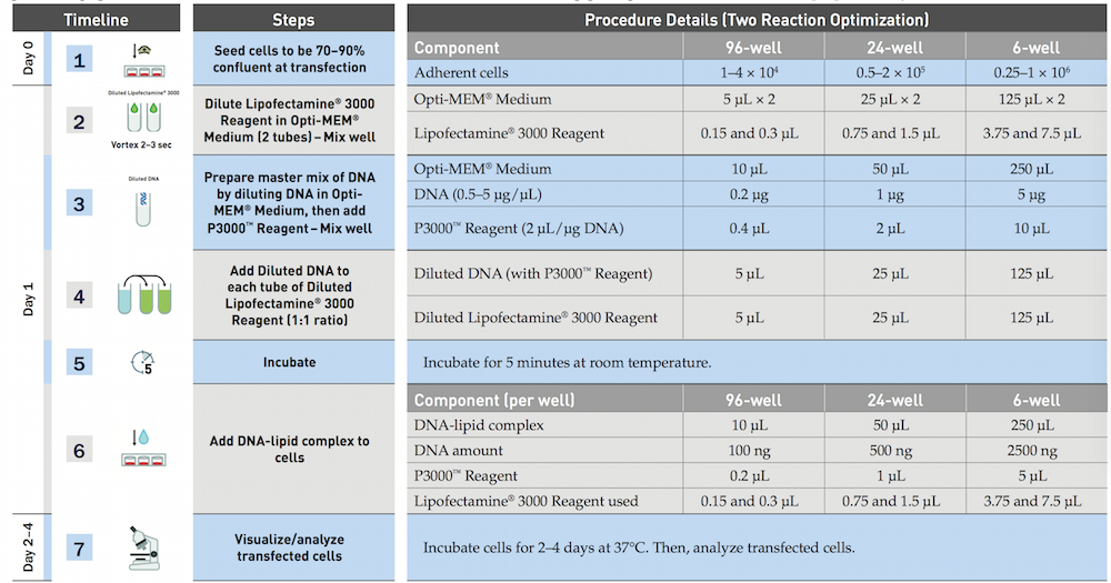
- Dilute 3.75μl/well Lipofectamine 3000 reagent in 25μl/well Opti-MEM, and incubation for 5min
- Prepare master mix of 5DNA μg/well by dilutiong DNA in Opti-MEM medium 25μl/well according to the form, then add P3000 reagent.
- Add dilutetd DNA to each tube of diluted lipo 3000.
- Incubation for 10min
- Add DNA lipid complex to cell waiting for the harvest.
Laboratory note
- Add DNA to 380μL Opti-MEM medium according to the table 2 and add 15μL P3000 reagent, mix well.
- Dilute Lipofectamine 3000 Reagent in Opti-MEM Medium: (2x) 1000μL Opti-MEM Medium + 120μL Lipofectamine 3000 Reagent, mix well.
- Add 395μL Diluted Lipofectamine 3000 to Diluted DNA of each group (1:1 ratio), mix well.
- Incubate for about 10min.
- Add DNA-lipid complex (250μL per well) to cells, shake the plate. (12:00 am)
- After 10h, change the medium with the complete medium with 10% FBS and Penicillin-Streptomycin.
Fluorescent Observation
[2014 Aug 16th 9:00am]
The first day after transfection
Change the medium of transfected cells and observe the fluorescent with fluorescent microscope.
Laboratory note=
The transfected cells are observed before we change the medium.
{SUSTC-Image|wiki/images/0/05/SUSTC-Shenzhen-3DayAfterTransefation-8.17G1.tif|G1 3day}
Figure 1.
//TODO figure
It is found that part of the cells round and suspended, and part of cells just become round. But the morphology of most of the cells are normal.
//TODO figure
Passage cell to dish
[2014 Aug 16th 15:00pm]
The first day after transfection
Passage part of cells to the 10cm petri dish (for picking monoclonal later except G5), and the remaining cells are still seeded on the previous 24-well plates (about 500μL cell suspension per well). (15:00 pm – 18:00 pm) For every group, two dishes with 5,000 cells seeded and two dished with 50,000 cells seeded.
Procedures
- Wash cells with PBS, trypsin digestion, add medium to stop digestion
- Transfer the cell suspension into 15 mL centrifuge tube and centrifuge at 800rpm for 5 min.
- Discard the supernatant and add 2 mL complete medium to suspend the cells.
- Count the cell concentration with blood counting chamber.
- Mark the new dishes, add 5 mL medium to every dish, and add corresponding cell suspension, mix well with the pipet.
- Culture.
| Group | Counted number | Concentration (cells/mL) | Volume needed |
|---|---|---|---|
| 1 | 30 | 300,000 | 167 and 16.7 |
| 2 | 20 | 200,000 | 294 and 29.4 |
| 3 | 30 | 300,000 | 294 and 29.4 |
| 4 | 30 | 300,000 | 143 and 14.3 |
Discussion
When counting cells, some dead cells were observed, and those cells were not included. The second day morning, some of the cells on the petri dish are round and suspended. Cells were few and scattered. For dishes with 5,000 cells, only several cells can be observed in one field under 4X objective lens. For dishes with 50,000 cells, near 10-20 cells can be observed in one field under 4X objective lens.
Fluorescent Observation
[2014 Aug 18th 10:00am]
The third day after transfection
Observe the transfected cells on the plate under fluorescence microscope.

Figure . The fluorescent picture of G1 captured by microscope. of Cells has been passage to dish so the concentration is low. The rate of transefaction is pretty high, because the usage of pBx-083 expressing EGFP is high.

Figure . The fluorescent picture of G2. Rate of fluorescent and intensity is much lower comparing with G1.

Figure . The fluorescent picture of G3. G3 has transfected with plasmid expressing Cas9 protein and GFP.

Figure . The fluorescent picture of G4. Same composition of plasmid as G3, but with lower usage of plasmid pBx-083 lower rate of transfection and lower intensity of fluorescencs.
[2014 Aug 18th 15:00pm]
The third day after transfection
And observe the transfected cells on petri dish with 50,000 cells under fluorescence microscope. For group 1 and 3 some living cells with fluorescence can be observed but for group 2&4, it is hard to find fluorescence. //TODO figure
Passage cells
[2014 Aug 19th 00:00am]
The forth day after transfection
Passage cells of every Groups from wells to petri dishes
Procedures
- Wash cells with PBS for 2 times
- Add trypsin 1ml and discard to digest for 2min at 37°C.
- Add complete medium 2ml to end digestion, suspend cells.
- Passage cell suspension to 10cm petri dish, and 5ml complete medium mix well with pipet. Mark the dish.
Cell selection
[2014 Aug 19th 16:00pm]
The forth day after transfection
Start the screening of the transfected cells on the plate with corresponding antibiotics.
Start the selection and screening of the transfected cells on one petri dish with 50,000 cells with corresponding antibiotics.
| Group | Blasticidin(ng/μL) | Puromycin(ng/μL) |
|---|---|---|
| 1&2 | 2.8 | 0 |
| 3&4 | 2.8 | 2 |
| 5 | 0 | 2 |
Fluorescent Observation
[2014 Aug 20th 18:00pm]
The fifth day after transfection
Observe the cells on the petri dish with fluorescence microscope. It is easy to find many living with green fluorescence in the pate, and the fluorescence of groups 1&3 is stronger than that of groups 2&4 moreover group 1&3 have much more cells with fluorescence than 2&4.
//TODO figure
[2014 Aug 22th 18:30pm]
The seventh day after transfection
Prepare for the second transfection. </br>
We think that the first transfection may not be good enough, as a result, we prepare for the second time transfection. Just the same as the first time, we schemed to transfect HeLa cells with 3 different combination of plasmid.
| Group A | EGFP:Piggybac = 1:1 |
|---|---|
| Group B | Cas9:EGFP:Piggybac = 1:1:1 |
| Group C | Cas9:Piggybac = 1:1 |
We seed HeLa cells in 6-well-plate (6wells at total), 100,000 cells per well. For tansfection
- Procedure
- Wash cells with PBS, trypsin digestion, and medium to stop digestion.
- Discard the supernatant and add 8ml complete medium to resuspend the cells.
- Add 1ml PBS to every gap between well
- 1:10 dilution count the cell concentration with blood counting slide. Get the concentration: 760,000 cells/ml. 0.78ml cell suspendion + about 9ml DMEM with 10% FBS. Mix well and add 1.5ml per well, to mix well pepite the well gently.
//TODO{
//TODO}
 "
"