Team:Lethbridge/results
From 2014.igem.org
| Line 82: | Line 82: | ||
</center> | </center> | ||
| - | <h2>Expression of Lamp2B construct | + | <h2>Expression and localization of RVG-Lamp2B protein construct</h2> |
| - | <p>< | + | <p>To test if our pcDNA3.0-RVG-Lamp2B construct was being appropriately expressed and folded, we fused Clover (a green fluorescent protein) to the C-terminus of Lamp2B. Using the Phyre2 server, we were able to generate a model of the predicted structure of our RVG-Lamp2B-Clover fusion protein based on homology and multi-template <i>ab initio</i> modeling.</p> |
| - | + | ||
<center>[[Image:Lamp2BClover.jpg|400px]]</center> | <center>[[Image:Lamp2BClover.jpg|400px]]</center> | ||
| - | <p><b>Figure 1 | + | <p><b>Figure 1: The predicted structure of the RVG-Lamp2B-Clover fusion protein.</b> The Clover domain is obvious in red with Lamp2B below in green and the RVG domain evident at the bottom in blue.</p><br> |
| - | <p> | + | <p>The pcDNA3.0-RVG-Lamp2B-Clover plasmid and a pcDNA3.0-Clover control were lipofected into separate Human Embryonic Kidney (HEK-293) cell cultures (HEK-293 cells are a resilient and rapidly dividing cell line also known to produce exosomes). In both lipofected cultures, there was elevated cytosolic green fluorescence relative to non-lipofected controls, which confirms expression and proper folding of the protein construct.</p> |
| - | <p> | + | <p>To confirm that the RVG-Lamp2B-Clover protein construct was being targeted to exosomal membranes (and were not simply cytosolic), culture media was collected from the lipofected and control HEK-293 cell cultures and exosomes were purified via ultracentrifugation. After lysis, the exosomes isolated from pcDNA3.0-RVG-Lamp2B-Clover lipofected cultures demonstrated an elevated level of fluorescence (505-620nm) that corresponds to the emission wavelength of Clover (515nm) indicating appropriate targeting of the engineered protein to exosomal membranes.</p> |
<center>[[Image:Fluorimeter_data.jpg|500px]]</center> | <center>[[Image:Fluorimeter_data.jpg|500px]]</center> | ||
| - | <p><b>Figure 2 | + | <p><b>Figure 2: Lysed exosomes isolated from pcDNA3.0-RVG-Lamp2B-Clover lipofected HEK-293 cells demonstrate elevated fluorescence in the 505-620nm range relative to control and unlysed samples.</p><br> |
<center>[[Image:CloverExosomes.jpg|300px]]</center> | <center>[[Image:CloverExosomes.jpg|300px]]</center> | ||
| - | <p><b>Figure | + | <p><b>Figure 3: Confocal Image of isolated exosomes carrying the Lamp2B-clover construct.</b> Description goes here.</p><br> |
Revision as of 00:20, 17 October 2014

Results
Team Parts Sandbox
Name Type Description Designer Length Favorite Part [http://parts.igem.org/Part:BBa_K1419000 BBa_K1419000] Device Arabinose inducible lysis casette Zak Stinson 3002 [http://parts.igem.org/Part:BBa_K1419001 BBa_K1419001] RNA Part Arabinose inducible lysis casette with RNA-IN Zak Stinson 32 [http://parts.igem.org/Part:BBa_K1419002 BBa_K1419002] Device RNA-OUT component for an Arabinose inducible lysis casette Harland Brandon 117****#$*#$ [http://parts.igem.org/Part:BBa_K1419003 BBa_K1419003] Device RNA-OUT component for a Universal Regulation System Harland Brandon 117****#$*#$ [http://parts.igem.org/Part:BBa_K1419004 BBa_K1419004] Device TEV Protease Harland Brandon 729DFDGFHFDHF [http://parts.igem.org/Part:BBa_K1419005 BBa_K1419005] Device Lamp2B-Clover Harland Brandon 2241 Yes Expression and localization of RVG-Lamp2B protein construct
To test if our pcDNA3.0-RVG-Lamp2B construct was being appropriately expressed and folded, we fused Clover (a green fluorescent protein) to the C-terminus of Lamp2B. Using the Phyre2 server, we were able to generate a model of the predicted structure of our RVG-Lamp2B-Clover fusion protein based on homology and multi-template ab initio modeling.
Figure 1: The predicted structure of the RVG-Lamp2B-Clover fusion protein. The Clover domain is obvious in red with Lamp2B below in green and the RVG domain evident at the bottom in blue.
The pcDNA3.0-RVG-Lamp2B-Clover plasmid and a pcDNA3.0-Clover control were lipofected into separate Human Embryonic Kidney (HEK-293) cell cultures (HEK-293 cells are a resilient and rapidly dividing cell line also known to produce exosomes). In both lipofected cultures, there was elevated cytosolic green fluorescence relative to non-lipofected controls, which confirms expression and proper folding of the protein construct.
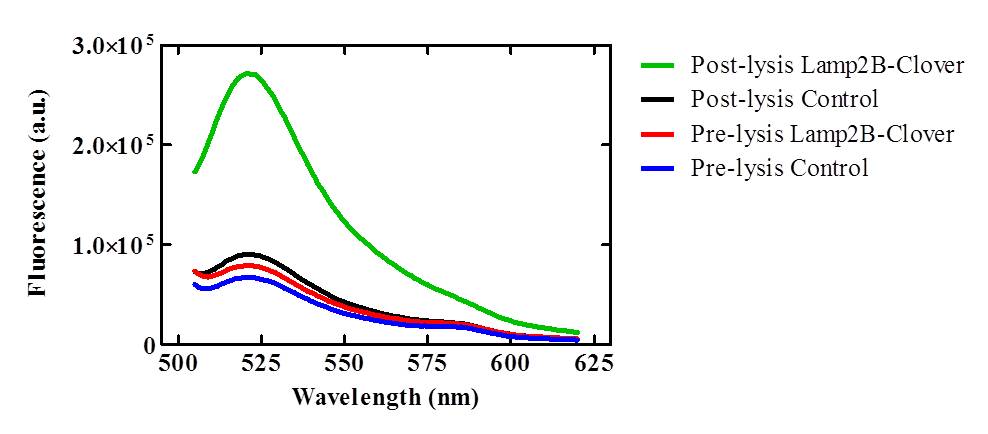
To confirm that the RVG-Lamp2B-Clover protein construct was being targeted to exosomal membranes (and were not simply cytosolic), culture media was collected from the lipofected and control HEK-293 cell cultures and exosomes were purified via ultracentrifugation. After lysis, the exosomes isolated from pcDNA3.0-RVG-Lamp2B-Clover lipofected cultures demonstrated an elevated level of fluorescence (505-620nm) that corresponds to the emission wavelength of Clover (515nm) indicating appropriate targeting of the engineered protein to exosomal membranes.
Figure 2: Lysed exosomes isolated from pcDNA3.0-RVG-Lamp2B-Clover lipofected HEK-293 cells demonstrate elevated fluorescence in the 505-620nm range relative to control and unlysed samples.</p>
<p><b>Figure 3: Confocal Image of isolated exosomes carrying the Lamp2B-clover construct. Description goes here.
Figure 1. Confocal Image of HEK cells transfected with Lamp2B-clover construct. Description goes here.
Figure 1. Confocal Image of HEK cells transfected with Lamp2B-clover construct. Description goes here.
 "
"


