Team:Austin Texas/photocage
From 2014.igem.org
Nathanshin (Talk | contribs) |
|||
| Line 113: | Line 113: | ||
*(+)IPTG, (+) ONBY | *(+)IPTG, (+) ONBY | ||
| - | Each of the conditions were in sterile test tubes and contained 1 mL of media and appropriate antibiotics. These cultures were allowed to grow for 2 hours. After 2 hours of growth, IPTG was added to all (+)IPTG condition tubes. The test tubes were allowed to grow another 2-4 hours, or until the OD600 reached 0.3. At this point, five 100 microliter samples of each of the strains were transferred into a 96-well plate. Each of the five samples represents a time interval of | + | Each of the conditions were in sterile test tubes and contained 1 mL of media and appropriate antibiotics. These cultures were allowed to grow for 2 hours. After 2 hours of growth, IPTG was added to all (+)IPTG condition tubes. The test tubes were allowed to grow another 2-4 hours, or until the OD600 reached at least 0.3. At this point, five 100 microliter samples of each of the strains were transferred into a 96-well plate. Each of the five samples represents a time interval of light exposure. |
| - | Next, the 96-well plate was covered in foil such that only the first column (which only contained samples that are to be | + | Next, the 96-well plate was covered in foil such that only the first column (which only contained samples that are to be exposed to 365 nm light for 30 minutes) was exposed. The handheld UV light was held 5cm over the 96 well plate and the first column was exposed to the 365 nm light for 15 minutes. After 15 minutes of exposure, the foil was removed to reveal the second column of cultures. Then, after another 10 minutes of exposure, the foil was removed to exposed the third column. After another 4 minutes, the fourth column was exposed. Finally, after the final 1 minute of exposure, the plate was re-covered with foil. After all of the sample were irradiated, an initial measurement of GFP expression was taken by the fluorometer. The purpose of the initial reading was to see what the initial effects of irradiation on the cultures were. |
Once the initial readings were recorded, the 100 microliters samples in the black 96 well plate were transferred into a deep 96 well plate, covered, and allowed to grow overnight at 37C and 225 RPM. | Once the initial readings were recorded, the 100 microliters samples in the black 96 well plate were transferred into a deep 96 well plate, covered, and allowed to grow overnight at 37C and 225 RPM. | ||
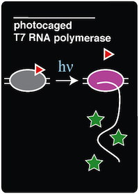
| - | The cultures were allowed 16 hours of growth so that the decaged T7 RNA polymerase would have time to polymerize mRNA transcripts of the GFP, which is | + | The cultures were allowed 16 hours of growth so that the decaged T7 RNA polymerase would have time to polymerize mRNA transcripts of the GFP, which is downstream of a T7 promoter. After 16 hours of growth, cultures were transferred back to a black 96 well plate and the fluorescence was measured. |
Revision as of 19:02, 17 October 2014
| |||||||||||||||||||||||||||||
 "
"


